Figures & data
Figure 1 Flow chart depicting the process for preparing the chitosan-coated collagen/β-TCP microspheres.
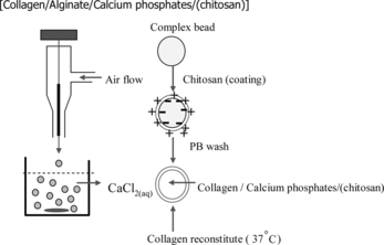
Figure 2 Removal of alginate from chitosan-coated collagen/β-TCP/alginate gel beads by PBS. The chitosan-coated collagen/b-TCP/alginate gel beads were immersed in PBS 37°C. The amount of the alginate releasing from the gel beads was measured according to the method described by Halle [Citation[10]].
![Figure 2 Removal of alginate from chitosan-coated collagen/β-TCP/alginate gel beads by PBS. The chitosan-coated collagen/b-TCP/alginate gel beads were immersed in PBS 37°C. The amount of the alginate releasing from the gel beads was measured according to the method described by Halle [Citation[10]].](/cms/asset/66bac487-e6c6-4925-9c70-498b9363c808/ianb19_a_237751_f0002_b.gif)
Figure 3 Optical micrograph of a chitosan-coated collagen/β-TCP microsphere. The black spots are due to the non transparent β-TCP particulates.
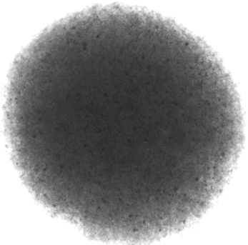
Figure 4 SEM micrograph of the chitosan-coated collagen/β-TCP microsphere: (a) surface (magnification: × 10000), and (b) inner section (magnification: × 10000). The microsphere contains 35% w collagen and 65% w β-TCP microsphere.
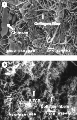
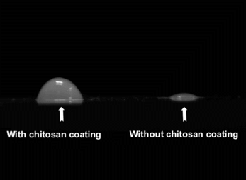
Figure 5 Optical micrographs of the microspheres on the glass slide: (left) microspheres with chitosan coating; (right) a microspheres without chitosan coating.