531
Views
17
CrossRef citations to date
0
Altmetric
Original

Rabbit Articular Chondrocytes Seeded on Collagen-Chitosan-GAG Scaffold for Cartilage Tissue Engineering In Vivo

, &
Pages 333-344 | Published online: 11 Jul 2009

Figures & data

Figure 1 The SEM photomicograph of Col-Chi-CS scaffold. A. The surface of scaffold. B. The cross-section of scaffold.
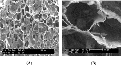
Figure 1 The SEM photomicograph of Col-Chi-CS scaffold. A. The surface of scaffold. B. The cross-section of scaffold.

Figure 2 Scanning electron micrographs chondrocytes seeded in Col-Chi-CS scaffold. A. Cell aggregates grew and adhered to scaffold after 3 days. B. Cells are surround by synthesized matrix after 21 days cell-seeding.
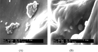
Figure 2 Scanning electron micrographs chondrocytes seeded in Col-Chi-CS scaffold. A. Cell aggregates grew and adhered to scaffold after 3 days. B. Cells are surround by synthesized matrix after 21 days cell-seeding.

Figure 3 H & E staining chondrocytes seeded in Col-Chi-CS matrix. A. Three days after seeding, × 100. B. Cells presented cartilage-specific lacunae in the superficial area 14 days after seeding, × 200. C. More homogeneous cartilage-like tissue were formed after 28 days seeding, × 100.
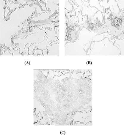
Figure 3 H & E staining chondrocytes seeded in Col-Chi-CS matrix. A. Three days after seeding, × 100. B. Cells presented cartilage-specific lacunae in the superficial area 14 days after seeding, × 200. C. More homogeneous cartilage-like tissue were formed after 28 days seeding, × 100.

Figure 4 A. Chondrocyte-scaffold constructs implanted subcutaneously in the back of athymic nude mouse. Photographs of mouse immediately after implantation after 12 weeks. B. The H&E staining of Chondrocytes/Col-Chi-CS scaffold implanted in vivo for 4 weeks (× 100); C. The H&E staining of Chondrocytes/Col-Chi-CS scaffold implanted in vivo for 12 weeks (× 100). D. The H&E staining of Chondrocytes/Col-Chi-CS scaffold implanted in vivo for 12 weeks (× 400).
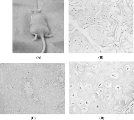
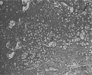
Figure 4 A. Chondrocyte-scaffold constructs implanted subcutaneously in the back of athymic nude mouse. Photographs of mouse immediately after implantation after 12 weeks. B. The H&E staining of Chondrocytes/Col-Chi-CS scaffold implanted in vivo for 4 weeks (× 100); C. The H&E staining of Chondrocytes/Col-Chi-CS scaffold implanted in vivo for 12 weeks (× 100). D. The H&E staining of Chondrocytes/Col-Chi-CS scaffold implanted in vivo for 12 weeks (× 400).

Figure 5 Toluidine blue staining of constructs confirmed chondorcytes abundance GAG after implanted in vivo for 12 weeks (× 100).

Figure 5 Toluidine blue staining of constructs confirmed chondorcytes abundance GAG after implanted in vivo for 12 weeks (× 100).

Reprints and Corporate Permissions

Please note: Selecting permissions does not provide access to the full text of the article, please see our help page How do I view content?

To request a reprint or corporate permissions for this article, please click on the relevant link below:

Academic Permissions

Please note: Selecting permissions does not provide access to the full text of the article, please see our help page How do I view content?

Obtain permissions instantly via Rightslink by clicking on the button below:

If you are unable to obtain permissions via Rightslink, please complete and submit this Permissions form. For more information, please visit our Permissions help page.