Figures & data
Table 1. Properties of PEG enzyme derivatives
Table 2. Storage stabilities of PEG enzyme derivatives at 4°C
Table 3. Encapsulation of PEG enzymes in erythrocytes
Table 4. Hemogram analysis of erythrocytes encapsulated at various urease/AlaDH activity ratios
Figure 1 Variation of average (a) blood urea, (b) blood ammonia values during the day in the dose control group injected with PEG enzyme preparation.
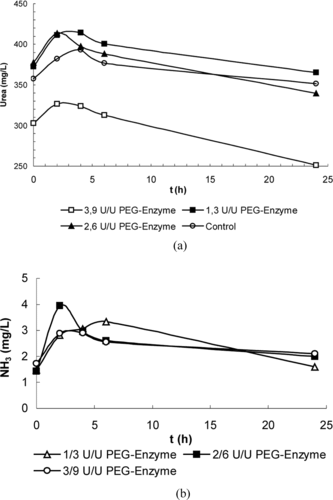
Table 5. Daily variations of the blood urea values in the control group
Figure 2 Daily variation of blood urea level in the PEG enzyme group.
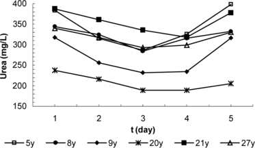
Figure 3 Daily variation of blood urea level in the erythrocyte group.
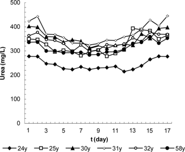
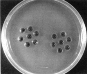
Figure 4 Precipitation test in agarose gel. 1/10 dilution of PEG-urease (left) and PEG-AlaDH (right) were applied into the central cavities; serum samples from 3 immunological groups and 3 control groups were applied to peripheric cavities.