425
Views
2
CrossRef citations to date
0
Altmetric
Original

Single Neural Progenitor Cells Derived from EGFP Expressing Mice is Useful After Spinal Cord Injury in Mice

, , &
Pages 405-414 | Published online: 11 Jul 2009

Figures & data

Figure 1 Neurosphere isolated from hippocampi of E-GFP transgenic mice (E13.5). A: All cells could express GFP, which is exhibiting green color under the excitation light. B: Cells were immunoreactive staining for nestin. C: Merged photo. The bar is 100 uM.
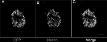
Figure 1 Neurosphere isolated from hippocampi of E-GFP transgenic mice (E13.5). A: All cells could express GFP, which is exhibiting green color under the excitation light. B: Cells were immunoreactive staining for nestin. C: Merged photo. The bar is 100 uM.

Table 1. Percentage of the glia and neuron cells after 7-day differentiation of EGFP NSCs in vitro

Figure 2 Representive photos for differentiation of EGFP-NSCs. A: GFAP positive cells. B: CNPase positive cells. C: MAP2 positive cells. D-F: Nuclei were stained with DAPI. G-I: Merged photos. The bar is 100 uM.
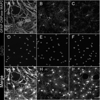
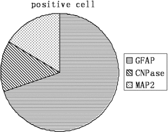
Figure 2 Representive photos for differentiation of EGFP-NSCs. A: GFAP positive cells. B: CNPase positive cells. C: MAP2 positive cells. D-F: Nuclei were stained with DAPI. G-I: Merged photos. The bar is 100 uM.
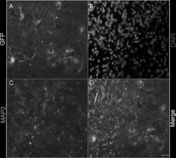
Figure 3 Percentage of the glia and neuron cells after 7-day differentiation of EGFP NSCs in vitro. A: : Percentage of the glia and neuron cells after 7-day differentiation of EGFP NSCs in vitro. B: The delineation of .

Figure 3 Percentage of the glia and neuron cells after 7-day differentiation of EGFP NSCs in vitro. A: Table 1: Percentage of the glia and neuron cells after 7-day differentiation of EGFP NSCs in vitro. B: The delineation of Table 1.

Figure 4 Differentiation of EGFP NSCs after transplantation into the leisured spinal cord in vivo. A: Transplantated EGFP positive cells. B: MAP2 positive cells. C: DAPI positive cells. D: Merged figure. Arrowheads denote double-labeled cells. The bar is 100 uM.

Figure 4 Differentiation of EGFP NSCs after transplantation into the leisured spinal cord in vivo. A: Transplantated EGFP positive cells. B: MAP2 positive cells. C: DAPI positive cells. D: Merged figure. Arrowheads denote double-labeled cells. The bar is 100 uM.

Reprints and Corporate Permissions

Please note: Selecting permissions does not provide access to the full text of the article, please see our help page How do I view content?

To request a reprint or corporate permissions for this article, please click on the relevant link below:

Academic Permissions

Please note: Selecting permissions does not provide access to the full text of the article, please see our help page How do I view content?

Obtain permissions instantly via Rightslink by clicking on the button below:

If you are unable to obtain permissions via Rightslink, please complete and submit this Permissions form. For more information, please visit our Permissions help page.