Figures & data
Figure 1 Analysis of the purity of purified porcine hemoglobin (A) and the molecular distribution of its glutaraldehyde-polymerized derivatives (B).
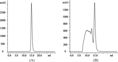
Figure 2 Silver-stained SDS-PAGE analysis of the purity of purified Hb and the molecular distribution of corresponding glutaraldehyde polymerized derivatives with 40 µg protein per lane (lane 1: pHb, lane 2: pPolyHb, lane 3: bHb, lane 4: bPolyHb, lane 5 :hHb and lane 6: hPolyHb).
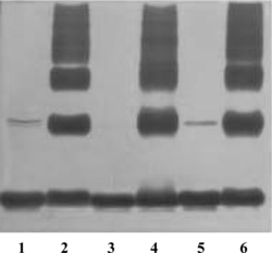
Table 1. Comparison of immunogenicity of different hemoglobin and their glutaraldehyde polymerized derivatives tested in mice
Figure 3 Western blotting analysis of cross-species binding reactions between a hemoglobin or its polymerized derivative and mouse polyclonal antibodies against a particular type of PolyHb.
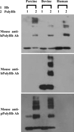
Table 2. Cross-species binding reaction (%) between glutaraldehyde polymerized hemoglobin and a particular ant-PolyHb polyclonal antibodies from rabbits or rats, as tested by a protein array method
Table 3. Amino acid sequence identity score among hemoglobin from different origins using ClustalW software for multiple alignment analysis