Figures & data
Scheme 1 Schematical description of “Separation Distance” measured by the AFM system.
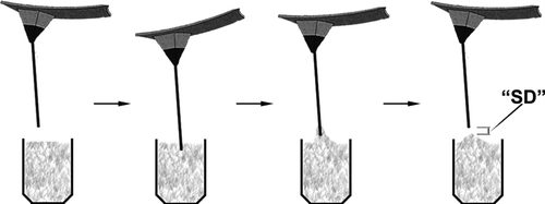
Figure 1 Representative optical micrographs of the AFM cantilever with 200X magnification (with 20X objective and 10X photoocular): (A) untreated; (B) after silanization step; and (C) after glutaraldehyde attachment. Pictures at the left column are the images without using filters, while the ones at the right column were after filtering by using Photoshop, glowing edges.
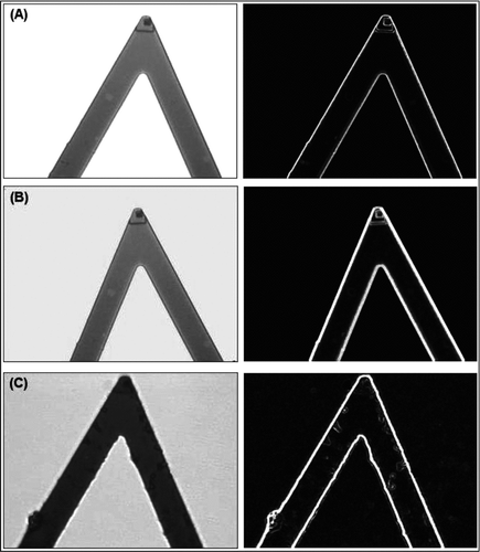
Figure 2 Representative confocal microscopy micrographs of the AFM cantilevers carrying histidine with 200X magnification (with 20X objective and 10X photoocular). Note that A, B, C are standard but D is a 3-D image of the AFM cantilever.
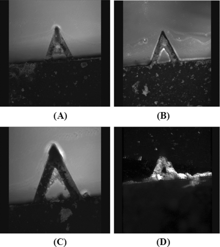
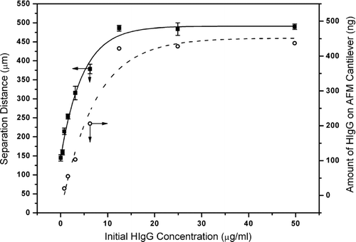
Figure 3 Change of separation distance (measured by the AFM system; average of ten repeated measurements with each five cantilevers and the standard deviation) and amount of HIgG adsorbed onto histidine carrying AFM cantilevers (obtained by the modified Lowry method).