Figures & data
Figure 1. Hepatic cells cultured on mineral fibers. A – Light microscopic picture of mineral fibers with hepatoma C3A cells. B – corresponding fluorescence picture with DAPI stained nuclei of human hepatoma C3A cells. C, D – Scanning electron microscopy pictures taken from human hepatoma C3A cells cultured on mineral fibers. C – human hepatoma C3A cell; F – mineral fiber.
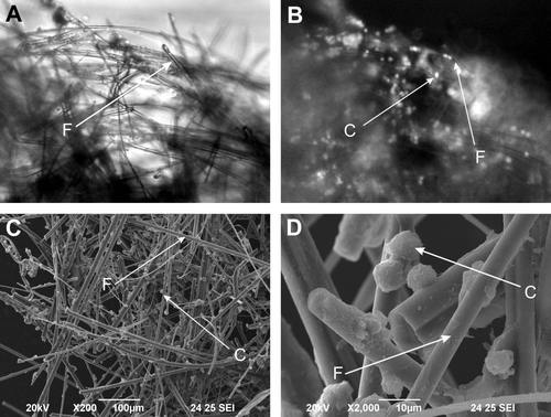
Figure 2. Total protein synthesis by human hepatoma C3A cells in bioreactor after 1, 6, and 24 hours of perfusion. Control – boioreactor perfused with control DMEM medium; PFC – bioreactor perfused with DMEM medium supplemented with 5% of PFC emulsion. (n = 6, *p < 0.05).
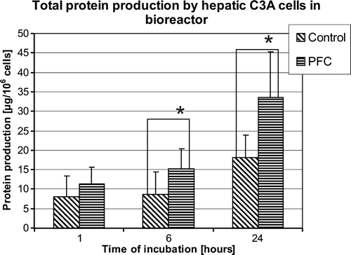
Figure 3. Albumin production by human hepatoma C3A in bioreactor after 1, 6, and 24 hours of perfusion. Control – bioreactors perfused with control DMEM medium; PFC – bioreactors perfused with DMEM medium supplemented with 5% of PFC emulsion. (n = 6, * p < 0.05).
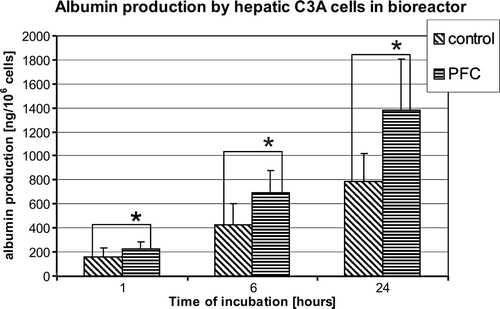
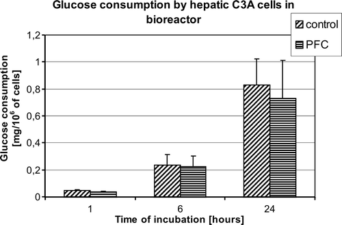
Figure 4. Glucose consumption by human hepatoma C3A cells in bioreactor after 1, 6, and 24 hours of perfusion. Control – bioreactor perfused with control DMEM medium; PFC – bioreactor perfused with DMEM medium supplemented with 5% of PFC emulsion.