Figures & data
Figure 1. The ideograph of biogradable conduit small gap bridging methods.
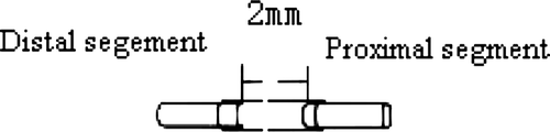
Figure 2. Conduit group S100 staining 10X.
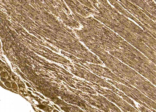
Figure 3. Conduit group HE staining 4X. Nerve fibers’ course in conduit 6 months operatively (longitudinal section).
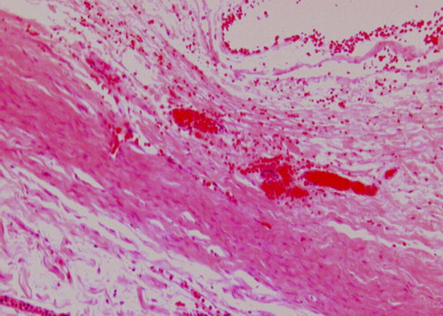
Figure 4. The course of nerve fibers in conduit 6 months operatively (cross section) 20X.
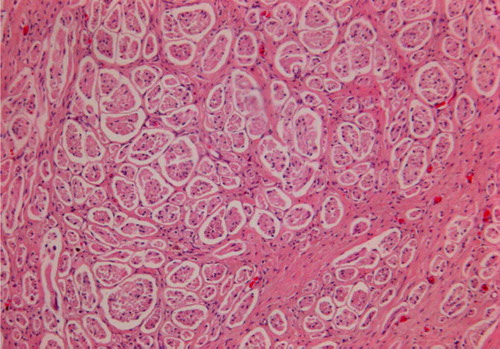
Figure 5. Regenerative nerve fiber in fasciculation in conduit group.
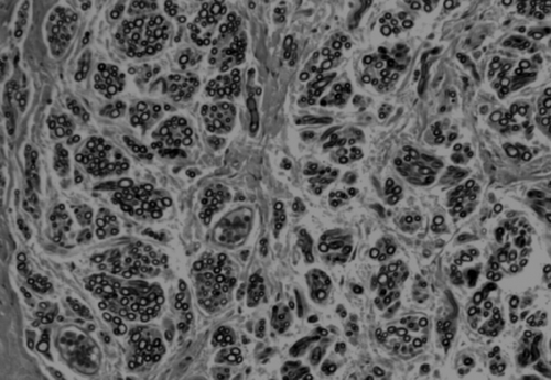
Figure 6. Regenerative nerve fiber in diffused distribution in traditionary epineurium suture group.
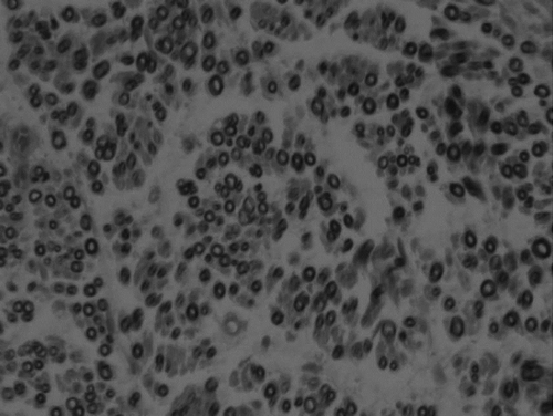
Table 1. Numbers of myelinated nerve fibers in all the experimental groups