Figures & data
Table 1. Diameter of the dermises
Figure 1. The moisture content of the dermises.
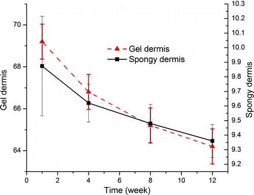
Table 2. The denature temperature of the dermises (°C)
Figure 2. Snap intensity of the dermises (Kpa).
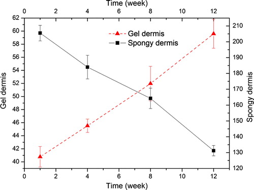
Figure 3. SEM of the dermises (A: spongy dermis; B: gel dermis).
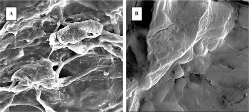
Figure 4. H&E staining of the dermises (A: spongy dermis cultured for 4 days; B: spongy dermis cultured for 8 days; C: gel dermis cultured for 4 days; D: gel dermis cultured for 4 days).
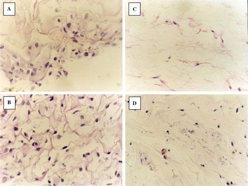
Table 3. The stains of the FN and type I collagen of the two scaffolds *
Figure 5. Phase contrast micrographs of primary cultured fibroblasts.
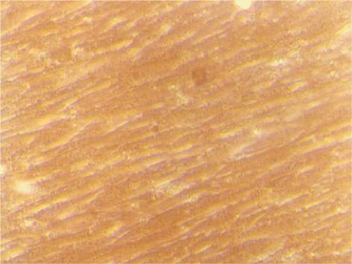
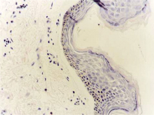
Figure 6. H&E staining of human skin.