117
Views
0
CrossRef citations to date
0
Altmetric
Erratum

Erratum

Pages 544-546 | Published online: 13 Aug 2009

Figures & data

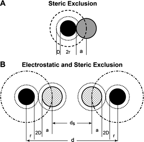
Appendix Figure A1. Diagram to illustrate steric plus electrostatic charge exclusion in a matrix. Radius of solute molecules (=a; radius of matrix molecules = r; D is the Debye length (the range of electrostatic influence) and this is ~1.0 nm in solutions the ionic strength of plasma. Figure A1A shows a neutral solute molecule (grey) adjacent to a charged matrix molecule of radius r (solid black). The centre of the neutral solute molecule is excluded from a region of solution of radius r + a (heavy broken line black distance a surrounding the matrix molecule but with no effect of the electrostatic exclusion zone a distance D (=1nm) surrounding the matrix (inner broken line). Figure A1B shows the case where negatively charged solutes (light hatched) are excluded by both steric and electrostatic charge (similar charge density on both solute and matrix). The center of the charged solute is excluded from a region of solution of radius r + a+2D (dot and dash line). The space available to 2 negatively charged solute molecules (ds) between matrix molecules that are a distance d apart when the matrix molecules also carry a negative charge is ds=d − 2(r + a+2D) whereas the space available to water and small solutes (a close to zero) = d − 2r.
Appendix Figure A1.  Diagram to illustrate steric plus electrostatic charge exclusion in a matrix. Radius of solute molecules (=a; radius of matrix molecules = r; D is the Debye length (the range of electrostatic influence) and this is ~1.0 nm in solutions the ionic strength of plasma. Figure A1A shows a neutral solute molecule (grey) adjacent to a charged matrix molecule of radius r (solid black). The centre of the neutral solute molecule is excluded from a region of solution of radius r + a (heavy broken line black distance a surrounding the matrix molecule but with no effect of the electrostatic exclusion zone a distance D (=1nm) surrounding the matrix (inner broken line). Figure A1B shows the case where negatively charged solutes (light hatched) are excluded by both steric and electrostatic charge (similar charge density on both solute and matrix). The center of the charged solute is excluded from a region of solution of radius r + a+2D (dot and dash line). The space available to 2 negatively charged solute molecules (ds) between matrix molecules that are a distance d apart when the matrix molecules also carry a negative charge is ds=d − 2(r + a+2D) whereas the space available to water and small solutes (a close to zero) = d − 2r.

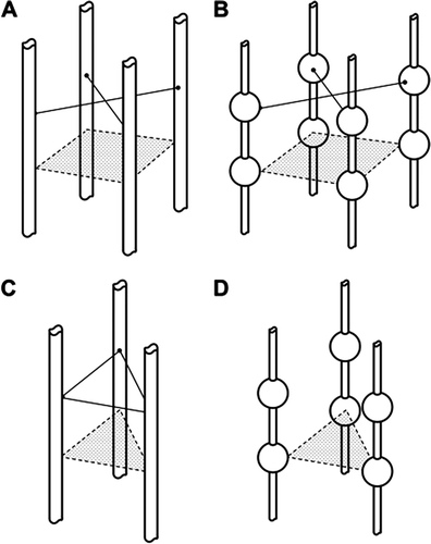
Appendix Figure A2. Four examples of possible structures for which λ has been calculated. Figs A2A and A2C represent the glycocalyx as a series of cylindrical molecular chains, which arise from trans-membrane proteins in the luminal endothelial cell membrane. In figures A2B and A2D the cylindrical chains link globular proteins (represented by spheres). In Figure 1A and 1B the chains are based on a square array, the diagonal of which is 11.2 nm (note the cylinders are further apart in A2C because the diagonal is measured between the spheres). In Figures A2C and A2D the chains arise from equilateral triangles of side length 11.2 nm. Calculations suggest that highest values of the partition coefficient (λ) between plasma and the fluid within the glycocalyx occur with variants of Figure A2D when λ may have a value as high as 0.4 and there is no cross linking. Cross-linking between the chains (shown as solid connecting lines) reduces λ to less than 0.05. For Figures A1A and A1C all values for λ are less than 0.05.
Appendix Figure A2.  Four examples of possible structures for which λ has been calculated. Figs A2A and A2C represent the glycocalyx as a series of cylindrical molecular chains, which arise from trans-membrane proteins in the luminal endothelial cell membrane. In figures A2B and A2D the cylindrical chains link globular proteins (represented by spheres). In Figure 1A and 1B the chains are based on a square array, the diagonal of which is 11.2 nm (note the cylinders are further apart in A2C because the diagonal is measured between the spheres). In Figures A2C and A2D the chains arise from equilateral triangles of side length 11.2 nm. Calculations suggest that highest values of the partition coefficient (λ) between plasma and the fluid within the glycocalyx occur with variants of Figure A2D when λ may have a value as high as 0.4 and there is no cross linking. Cross-linking between the chains (shown as solid connecting lines) reduces λ to less than 0.05. For Figures A1A and A1C all values for λ are less than 0.05.

Reprints and Corporate Permissions

Please note: Selecting permissions does not provide access to the full text of the article, please see our help page How do I view content?

To request a reprint or corporate permissions for this article, please click on the relevant link below:

Academic Permissions

Please note: Selecting permissions does not provide access to the full text of the article, please see our help page How do I view content?

Obtain permissions instantly via Rightslink by clicking on the button below:

If you are unable to obtain permissions via Rightslink, please complete and submit this Permissions form. For more information, please visit our Permissions help page.