
Figures & data
Figure 1. Presentation of marker setup.
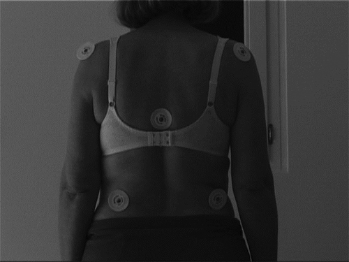
Table 1. The mean (95% confidence intervals) of asymmetry indices of weight distribution (AIW) and trunk angles (AIA) in healthy subjects and stroke patients.
Figure 2A. Relative ground force (RGF)in starting position (SP) in patients and controls.
*P < 0.05 compared to control group. #P < 0.05 compared to contralateral side.
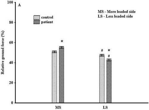
Figure 2B. Relative ground force (RGF) in weight shift position in patients and controls.
*P < 0.05 compared to control group. #P < 0.05 compared to contralateral side.
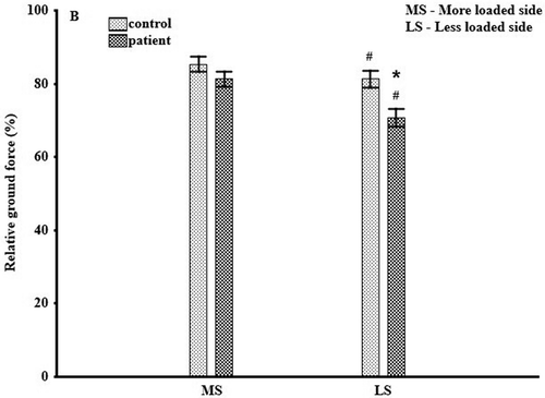
Figure 3. Trunk angles (AVIAs) on both sides in starting and weight shift positions in patients and controls. *P < 0.05 compared to control group. #P < 0.05 compared to contralateral side (MS: more loaded side; LS: less loaded side WS: weight shift).
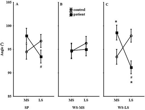
Figure 4. Elongation indices on both sides in starting and weight shift positions in patients and controls.*P < 0.05 compared to control group. #P < 0.05 compared to contralateral side (MS: more loaded side/LS: less loaded side; WBS: weight-bearing side; non-weight-bearing side).
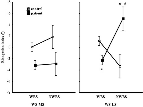
Figure 5. Changes of trunk alignment during weight shift in a healthy control subject (top) and a stroke patient (bottom). SP: The starting position (quiet standing); L: shift to the left; R: shift to the right.
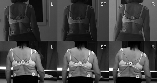
Table 2. Correlations between relative ground force (RGF) and acromion–vertebra–iliac crest angle (AVIA) values.
Table 3. Correlations between the asymmetry/elongation indices.