

Abstract
In the present study, the cellulose from sugarcane tops (SCT) was separated and characterized for its purity. Approximately, 85% (w/w) of total cellulose present in raw SCT was recovered by using alkaline method. The monosaccharide analysis of SCT cellulose by HPLC showed 91% D-glucose, 7.5% D-xylose and 1.5% D-arabinose residues. Surface morphology study of dried cellulosic fibers by FESEM exhibited the fibrous structure. The FTIR analysis of separated cellulose displayed the peaks corresponding to the peaks obtained from commercial cellulose, confirming its purity. The crystallinity index (CrI) of separated cellulose increased to 49% after delignification and xylan extraction from 36% of raw SCT. The typical TGA curve of separated SCT cellulose showed decomposition and mass reduction at 327 °C resulting in single decomposition peak in TGA analysis, confirming its purity. CHNS analysis supported the purity of separated cellulose by confirming absence of nitrogen and sulfur. The separated cellulose was hydrolyzed by recombinant endo-β-1,4-glucanase (CtCel8A), cellobiohydrolase (CtCBH5A) from Clostridium themocellum and β-1,4-glucosidase (HtBgl) from Hungateiclostridium thermocellum at pH 5.8, 50 °C for 24 h, resulting in the production of 188 mg/g of total reducing sugar (TRS). The separated cellulose from SCT can be utilized as an alternative substrate for commercialization and for bioethanol production.
GRAPHICAL ABSTRACT
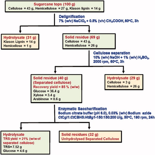
85% of total cellulose recovery yield was obtained from raw sugarcane top (SCT)
SCT cellulose contains 91%, D-glucose, 7.5% D-xylose and 1.5% D-arabinose residues
Crystallinity index (CrI) of separated cellulose increased from 36% to 49% of raw SCT
TGA curve of separated SCT cellulose showed the single decomposition peak at 327 °C
Separated cellulose hydrolyzed by recombinant enzymes gave 188 mg/g TRS yield
Highlights
Disclosure statement
The authors do not have any potential conflict of interest.
Acknowledgments
The authors acknowledge the use of FTIR spectrophotometer procured through the Indo-Finnish project grant (BT/IN/Finland/08/AG/2011) from Department of Biotechnology (DBT), Ministry of Science and Technology, Government of India to AG. The authors acknowledge the Central Instrument Facility (CIF) at Indian Institute of Technology (IIT), Guwahati, for the provision of FESEM, XRD and TGA facilities.