Figures & data
Fig. 1 Illustrative photo of the CE-MS system.
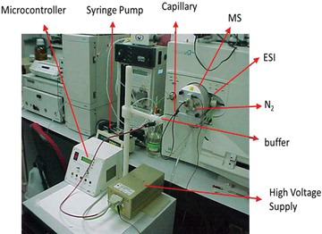
Table 1. Voltage regulators (Texas Instruments)
Fig. 2 The baseline of the CE–MS system was obtained by injecting the background electrolyte (BGE) under the following conditions: 1% acetic acid, 50% acetonitrile, 49% water; ESI voltage: 4.5 kV, and ESI temperature: 200°C. The auxiliary liquid was the same as BGE. The silica capillary was 50 cm in length, and E = 350 V cm−1. Samples were injected hydrodynamically for 5 s by raising the silica capillary injection end 10 cm above the level of the detection end.
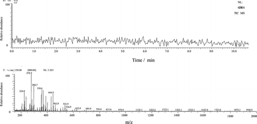
Fig. 3 Electropherogram/mass spectra. Sample: aprotinin 5.0 nmol µL−1 Other conditions were identical to those shown in Figure .
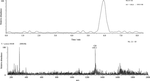
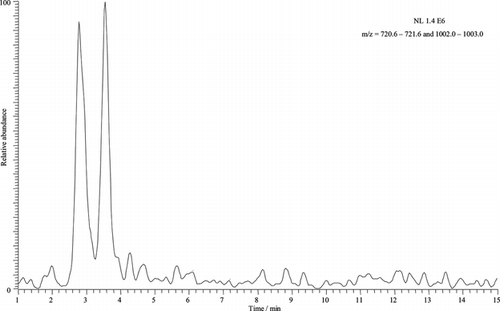
Fig. 4 Electropherogram/spectrum of BSA 5.0 nmol µL−1, buffer 0.5% acetic acid in water, E = 200 V cm−1, pH 3.5, ESI voltage: 4.5 kV, ESI temperature: 200°C. Sampling was performed in the same conditions as those shown in Figure .
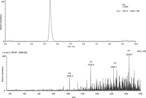
Fig. 5 Electropherogram obtained in a sheath-flow CE-MS system of lysozyme and BSA respectively. Separation electrolyte 1.0% acetic acid and PDMA capillary. E = 200 V cm−1, pH 3.5, ESI voltage: 4.5 kV, ESI temperature: 200°C. The sampling procedure was the same as that shown in Figure .