
Figures & data
Figure 1. Smartphone ophthalmoscope.
Note: A. Light-emitting diode in close proximity to the camera lens on the smartphone as almost co-axial illumination. B. USB cable with a corresponding connector (Type-C, Micro and Lightning from left to right). C. Screen capture of the camera app (Ullman Indirect) in the smartphone.
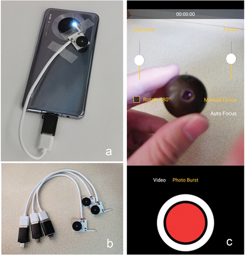
Figure 2. Study design.
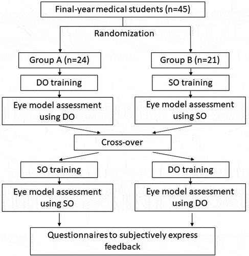
Figure 3. Trainings of ophthalmoscopy using smartphone ophthalmoscopes.
Note: Demonstration and practice using the smartphone ophthalmoscope on patients in the darkroom (A). Fundus image captured with smartphone ophthalmoscope on patients with undilated (B) and dilated (C) pupils. Written informed consents had been obtained for the image to be published.
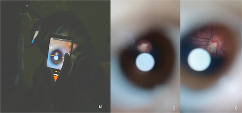
Figure 4. Students using ophthalmoscopes to visualize the fundus of the eye model.
Note: A and B. Visualizing the fundus of eye model using smartphone ophthalmoscope. C. Image captured using the smartphone ophthalmoscope. D. Visualizing the fundus of eye model using a direct ophthalmoscope.
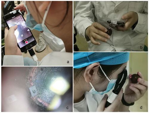
Table 1. Baseline characteristics of the study population. Data presented as means ± SD or numbers.
Table 2. Objective assessment of ophthalmoscopy.
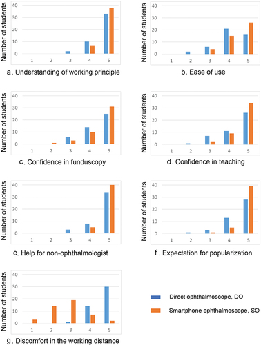
Figure 5. Subjective assessment of ophthalmoscopy using DO and SO .
Note: Number of students who provided a rating from 1 to 5
Data availability statement
All data analyzed or generated during the study are available from the corresponding author on reasonable request.