
Figures & data
Figure 1. Experimental design and procedures used to assess bicep femoris long head fascicle length. A. Image acquisition using 10-cm ultrasound probe, with probe-oriented perpendicular to the skin following the line of the bicep femoris (ischial tuberosity to lateral epicondyle). B. Experimental design with a timeline of test occasions and image acquisition. C. Example of sonogram image obtained of the bicep femoris with architectural features identified (muscle thickness, pennation angle, fascicle and aponeuroses (deep and superficial)).
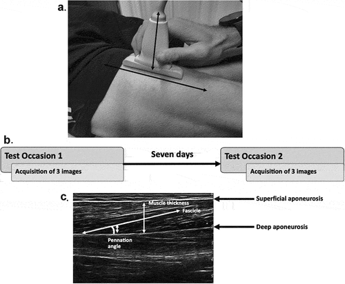
Table 1. Between-session mean (SD), reliability and error statistics for bicep femoris long head architectural measurements.
Figure 2. Differences in estimated fascicle length between the three methods of estimation, * = significant difference (p <0.05). Black line signifying mean estimated fascicle length, where circles signify individual measurements.
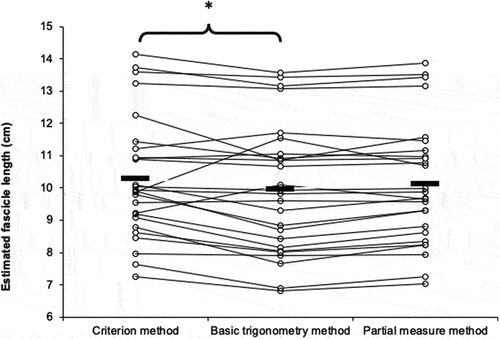
Figure 3. Bland Altman plots comparing the mean estimated fascicle lengths between methods. A) criterion vs. basic trigonometry and B) criterion vs. partial measure methods.
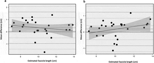
Table 2. Bias and limits of agreement between the estimated measures of bicep femoris fascicle length.
Figure 4. Relationship and 95% confidence limits between the criterion and alternative methods of estimating bicep femoris long head fascicle length.
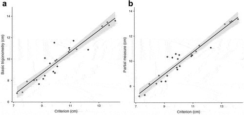
Table 3. Observed relationships between the estimated measures of bicep femoris fascicle length.