Figures & data
Figure 1. Geological map of Skåne (southern Sweden) with studied localities, modified after Kruger et al. (Citation2021, this issue). 1. Åbro BH-207/64 and Billesholm. 2. Hyllinge. 3. Textile Factory, Eslöv. 4. Höllviken II. 5. Stabbarp BH-74. 6. Helsingborg harbour. 7. Svedala I. 8. Kävlinge BH-928
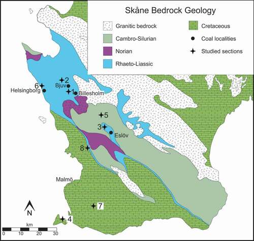
Figure 2. Early Mesozoic megaspore assemblages recorded in this study from Skåne (Sweden) and Bornholm (Denmark). G. Galgeløkke Member
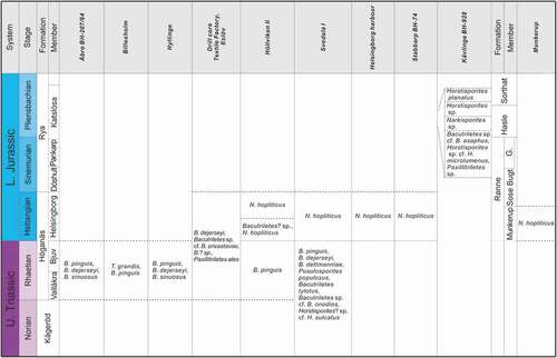
Figure 3. Stratigraphical occurrences of Triassic and Jurassic megaspores recorded in Skåne and Bornholm
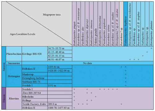
Figure 4. Trileites and Banksisporites. Scale bars = 100 μm. A, B. Trileites grandis Fuglewicz, Citation1973. A. Proximal view, SEM, S054719-02-01. B. Proximal view, SEM, S054353-03. C–F. Banksisporites dettmanniae Banerji, Kumaran et Maheshwari Citation1978. C. Proximal view, transmitted light, S079701, R36. D. Proximal view, transmitted light, overexposure, S079701, R36. E. Proximal view, fluorescence, S079701, R36. F. Distal view, fluorescence, S079701, R36
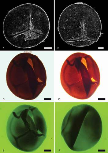
Figure 5. Banksisporites. Scale bars = 100 μm. A–F. Banksisporites pinguis (Harris, Citation1935) Dettmann, Citation1961. A. Proximal view, transmitted light, S054802-01-03, R40-2. B. Proximal view, fluorescence, S054802-01-03, R40-2. C. Megaspore tetrad, transmitted light, S064827-02-01, F31-2. D. Megaspore tetrad, fluorescence, S064827-02-01, F31-2. E. Lateral view, transmitted light, S054802-01-04, P38-3. F. Lateral view, fluorescence, S054802-01-04, P38-3. G–H. Banksisporites sinuosus Dettmann, Citation1961. G. Proximal view, transmitted light, S054802-05-01, R37. H. Proximal view, fluorescence, S054802-05-01, R37. I–L. Banksisporites dejerseyi Scott et Playford, Citation1985. I. Oblique view, transmitted light, S064850-02-01, P29-1. J. Oblique view, fluorescence, S064850-02-01, P29-1. K. Proximal view, transmitted light, S054802-05-01, R37. L. Proximal view, fluorescence, S054802-05-01, R37
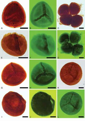
Figure 6. Pusulosporites populosus Fuglewicz, Citation1973. Scale bars = 100 μm. A. Distal view, transmitted light, S079726, O53-2. B. Distal view, fluorescence, S079726, O53-2. C. Proximal view, transmitted light, S079727, M43. D. Proximal view, fluorescence, S079727, M43. E. Proximal view, transmitted light, S079716, L37. F. Proximal view, fluorescence, S079716, L37
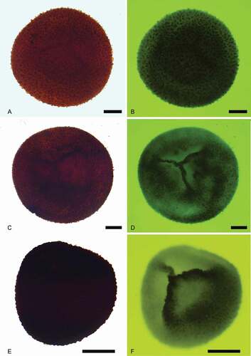
Figure 7. Bacutriletes tylotus (Harris, Citation1935) Potonié, Citation1956. Scale bars = 100 μm. A. Proximal view, transmitted light, S079701, R37-1. B. Proximal view, fluorescence, S079701, R37-1. C. Lateral view, transmitted light, S079701, V38. D. Lateral view, fluorescence, S079701, V38. E. Distal view, transmitted light, S079701, J37-2. F. Distal view, fluorescence, S079701, J37-2
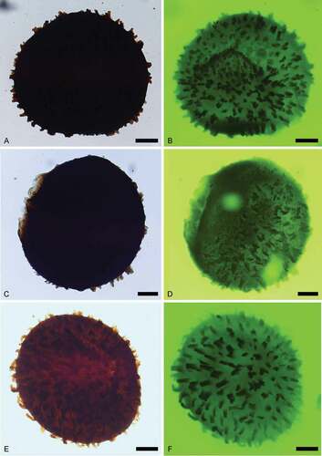
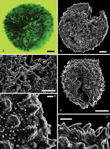
Figure 8. Narkisporites, Bacutriletes and Horstisporites. Scale bars = 100 μm, or indicated otherwise. A, B. Narkisporites sp. A. Lateral view, SEM, Kävlinge BH-928, 42.25–41.15 m. B. Enlargement, SEM, showing blunt and spiny projections outside the contact area, Kävlinge BH-928 42.25–41.15 m. Scale bar = 50 μm. C, D. Bacutriletes sp. cf. B. asaphus Fuglewicz, Citation1973. C. Proximal view, SEM, Kävlinge BH-928, 46.91–46.07 m. D. Enlargement, SEM, Kävlinge BH-928, 46.91–46.07 m. Scale bar = 50 μm. E, F. Horstisporites sp. cf. H. microlumenus Dettmann, Citation1961. E. Proximal view, SEM, Kävlinge BH-928, 46.91–46.07 m. F. Enlargement, SEM, Kävlinge BH-928, 46.91–46.07 m. Scale bar = 50 μm
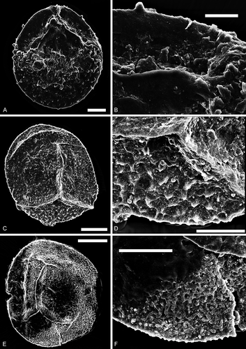
Figure 9. Bacutriletes. Scale bars = 100 μm. A–F, I, L. Bacutriletes? sp. A. Oblique view, transmitted light, S066549, R26. B. Oblique view, transmitted light, over-exposure, S066549, R26. C. Oblique view, fluorescence, S066549, R26. D. Proximal view, transmitted light, S063493-02, N42-4. E. Proximal view, fluorescence, S063493-02, N42-4. F. Enlargement of C, fluorescence, S066549, R26. I. Enlargement of C, fluorescence, S066549, R26. L. Distal view of a broken specimen, fluorescence, S063493-01, N42-4. G, H. Bacutriletes sp. cf. B. srivastavae Banerji, Jana et Maheshwari Citation1984. G. Lateral view, transmitted light, S063489, M44-4. H. Lateral view, fluorescence, S063489, M44-4. J, K. Bacutriletessp. cf. B. onodios (Harris, Citation1961) Hopkins et Sweet, Citation1976. J. Proximal view, fluorescence, S079718-01, P40-3. K. Distal view, fluorescence, S079718-01, P40-3
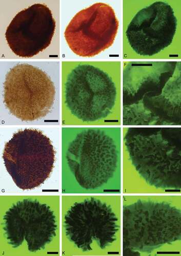
Figure 10. Horstisporites. Scale bars = 100 μm, or indicated otherwise. A–D. Horstisporites planatus (Marcinkiewicz, Citation1960) Marcinkiewicz, Citation1971. A. Proximal view, SEM, Kävlinge BH-928, 34.75–33.75 m. B. Enlargement, SEM, showing the micro-reticulation in the surface reticulation, Kävlinge BH-928, 34.75–33.75 m. Scale bar = 20 μm. C. Proximal view, SEM, Kävlinge BH-928, 34.75–33.75 m. D. Enlargement, SEM, showing the micro-reticulation in the surface reticulation, Kävlinge BH-928, 34.75–33.75 m. Scale bar = 10 μm. E, F. Horstisporites sp. E. Oblique view, SEM, Kävlinge BH-928, 41.15–40.40. F. Enlargement, SEM, Kävlinge BH-928, 41.15–40.40. Scale bar = 50 μm
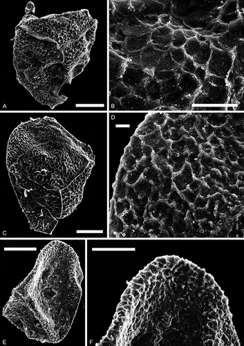
Figure 11. Horstisporites? sp. cf. H. sulcatus Fuglewicz, Citation1973. Scale bars = 100 μm. A. Oblique view, transmitted light, S079713, D31-3. B. Oblique view, fluorescence, S079713, D31-3. C. Lateral view, transmitted light, S079725, K34-3. D. Lateral view, fluorescence, S079725, K34-3. E. Distal view, transmitted light, S079701, L32-2. F. Distal view, fluorescence, S079701, L32-2
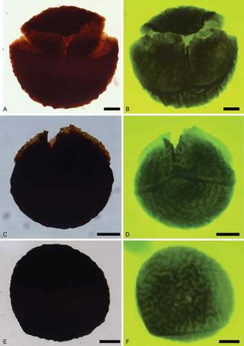
Figure 12. Paxillitriletes. Scale bars = 100 μm, or indicated otherwise. A, B. Paxillitriletes ales (Harris, Citation1935) Batten et Koppelhus, Citation1993. A. Lateral view, transmitted light, S063488, M42-3. B. Lateral view, fluorescence, showing strongly developed capilli near triradiate lamellae, S063488, M42-3. C–F. Paxillitriletes sp. C. Lateral view, SEM, Kävlinge BH-928, 46.91–46.07 m. D. Enlargement, SEM, Kävlinge BH-928, 46.91–46.07 m. Scale bar = 30 μm. E. Enlargement, SEM, Kävlinge BH-928, 46.91–46.07 m. Scale bar = 50 μm. F. Enlargement, SEM, Kävlinge BH-928, 46.91–46.07 m. Scale bar = 50 μm
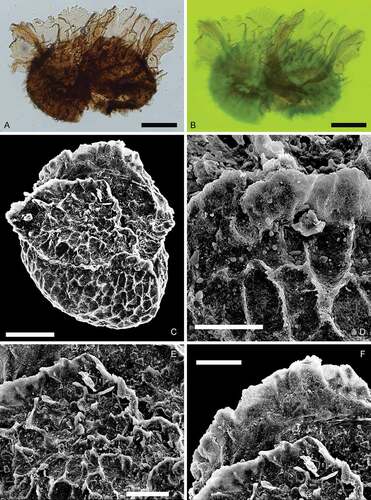
Figure 13. Nathorstisporites hopliticus Jung, Citation1958. Scale bars = 100 μm. A. Oblique view, transmitted light, S079741, L41-4. B. Oblique view, fluorescence, S079741, L41-4. C. Enlargement, fluorescence, S079741, L41-4. D. Enlargement, fluorescence, S079741, L41-4. E. Lateral view, transmitted light, S075612, M38-2. F. Lateral view, fluorescence, S075612, M38-2
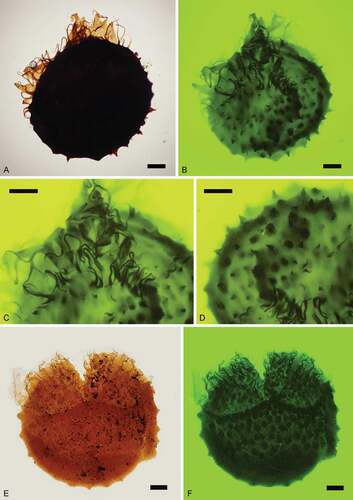
Figure 14. Nathorstisporites hopliticus Jung, Citation1958. Scale bars = 100 μm, or indicated otherwise. A. Proximal view, fluorescence, S066557, P36-2. B. Proximal view, SEM, Munkerup. C. Enlargement, SEM, Munkerup. Scale bar = 50 μm. D. Distal view, SEM, Munkerup. E. Enlargement, SEM, Munkerup. Scale bar = 10 μm. F. Enlargement, SEM, Munkerup. Scale bar = 20 μm