Figures & data
Figure 1. Locality information for GGU sample 216842 Cape Schuchert Formation (Silurian, Llandovery Series), Washington Land, North Greenland.
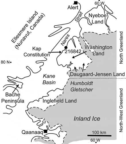
Figure 2. Holdfasts of Sphenothallus sp., GGU sample 216842 Cape Schuchert Formation (Silurian, Llandovery Series), Washington Land, North Greenland. A–C. PMU 38328/1, basal view of the holdfast cone with margin of basal attachment disc preserved at periphery (A and inset). D,E. PMU 38328/2, basal view of holdfast cone (E) showing dense, thin, outer layer and amorphous inner layer; detail in (D). F,G. PMU 38328/3, lateral views showing growth displacement (arrows). H,I. PMU 38328/4, holdfast in plan and oblique views. J. PMU 38328/5, oblique lateral view showing growth lamellae (arrow). K. PMU 38328/6, lateral view. L. PMU 38328/7, lateral view. M. PMU 38328/8, lateral view with irregular holdfast. N. PMU 38328/9, broken tube showing thickening of elliptical shell in apertural view. O. PMU 38328/10, oblique view, showing thickening of shell wall. P. PMU 38328/11, lateral view showing preferential breakage of lateral area. Scale bars: 20 µm (A); 50 µm (D,E); 100 µm (B,C,I,J,O); 200 µm (F–H,K–N,P).
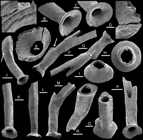
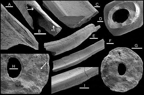
Figure 3. Sphenothallus sp., GGU sample 216842, Cape Schuchert Formation (Silurian, Llandovery Series), Washington Land, North Greenland. A,B PMU 38328/11, growth lamellae, with A a detail of B, the latter showing decrease in thickness of lamella from arrow a to arrow b. C,F,I. PMU 38328/12, channel-like fragment of longitudinal thickening showing growth lamellae. D. PMU 38328/10, apertural view of specimen illustrated as Fig. 2O showing thickening of shell wall and comarginal growth lamellae. E. PMU 38328/13, fragment of tube showing delimitation of longitudinal thickenings by groove (aperture to left). G,H. PMU 38328/14, inner surface of basal attachment disc with down-stepping growth lamellae (arrow in H). Scale bars: 50 µ m (A,C,D,H); 100 µm (B,G,I); 200 µm (E,F).