Figures & data
Figure 1. Maps showing locality where the sample was collected (star in C). The shaded area in Fig. A shows the County of Västerbotten. The sample was found ca. 560 m NNE of Jillesnåle farm, situated close to the northern shore line of the northwestern end of Lake Storvindeln (Fig. C) in northwestern Västerbotten County, Sweden.
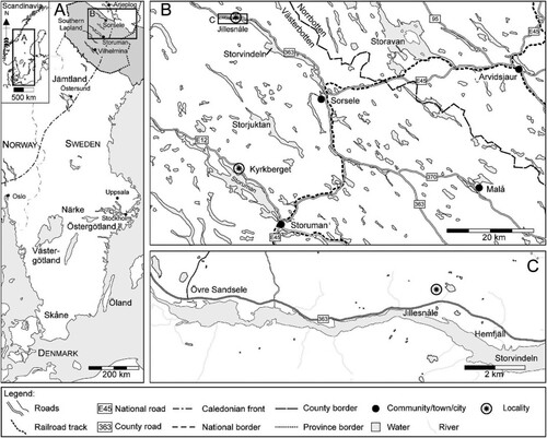
Figure 2. Simplified correlation chart of the Tremadocian strata of Norway and Sweden. The references to geographical directions with each locality column indicate the areas relative position on the Baltoscandian platform. Sedimentologically the western carbonate/glauconite facies represent distal ramp environments while the eastern are proximal ramp environments (Egenhoff et al. Citation2010). The star in the last column indicates inferred position of the sample from Jillesnåle. Stage Slices and conodont zonations are largely based on Bergström et al. (Citation2009). See text for further references.
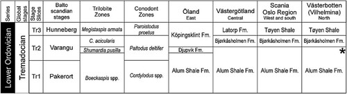
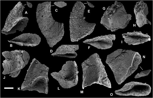
Figure 3. A–E. Mytoella? n. sp. A–C. Ventral valve (PMU38359) in external, internal and lateral view. D, E. Shell fragments PMU38361 and PMU38376 in external and internal view, respectively. F. Obolinae gen. and sp. indet. (PMU38385), exterior of exfoliated ventral valve. G. Thysanotos? sp. (PMU38365), recrystallized shell fragment with marginal spines. Scale bar equals 100 µm, except G (200 µm).
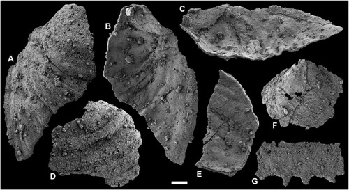
Figure 4. A, B, D. Eurytreta cf. sabrinae (Callaway, Citation1877). A, D. Ventral valve (PMU38362) in oblique anterior and lateral view showing apical process and broad vascula lateralia. B. Ventral valve (PMU38363) in posterior view showing larval shell and flat undivided pseudointerarea. C, F, G. Two dorsal valves of aff. Eurytreta sp. (C, PMU38374 and F, G, PMU38375) showing subrectangular median buttress and broad triangular median groove. E, H, I. Ottenbyella sp. (PMU38364) in ventral exterior, interior, and lateral view; note subrectangular apical process and narrow vascula lateralia. J, K, Dorsal valve of aff. Acrotreta sp. (PMU38360) with triangular median septum. L. Potential ventral valve of aff. Acrotreta sp. (PMU38386). Scale bar equals 100 µm.
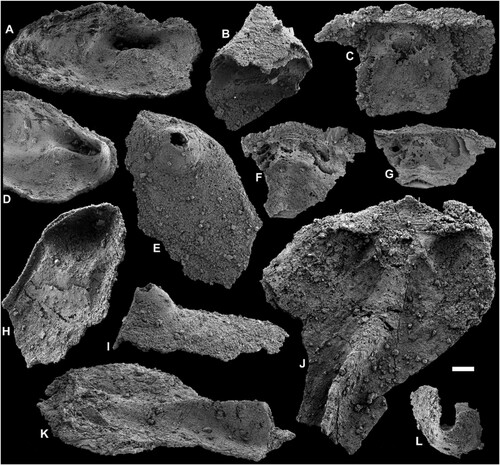
Figure 5. A–J, L–N. Pomeraniotreta biernatae Bednarczyk, Citation1986; A, ventral valve (PMU38380), posterior view showing undivided pseudointerarea; B, ventral valve (PMU38377), anterior view; C, ventral valve (PMU38379), oblique lateral view; D, E, apical portions of two ventral valves (PMU38367, PMU38384) showing profile of larval shell (for better display purposes E has been mirrored digitally); F, ventral valve (PMU38378), internal view showing apical process and pedicle tube; G, dorsal valves (PMU38368) with transversely oval larval shell; H, slightly concave dorsal valve (PMU38370) in lateral view, I, dorsal valve (PMU38373) with elongate oval larval shell; J, apical view of D; L, dorsal view of H; M, dorsal valve (PMU38371) interior view with subrectangular median buttress and faint interridge; N, dorsal valve (PMU38372), secondarily deformed and elongated. K, O, P, Ventral valve of Pomeraniotreta n. sp. (PMU38387) in posterior, right lateral, and left lateral view; note faint intertrough in K. Scale bar equals 100 µm.
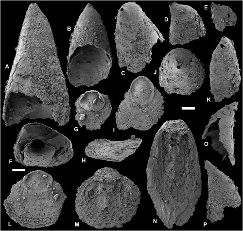
Figure 6. A, B, Ventral and dorsal larval shell of Pomeraniotreta biernatae (PMU38366, PMU38370). C, Ventral larval shell of Pomeraniotreta n. sp. (PMU38387, same specimen as in K, O, P). D, Columnar shell structure of P. biernatae seen in a dorsal valve (PMU38369). E, Anterior view of exfoliated ventral valve of P. biernatae exposing thick laminae of apical process (PMU38378, same specimen as in F). F, Larval shell pitting in a dorsal valve of P. biernatae (PMU38373, same specimen as in I). G, Larval shell pitting in a ventral valve of Pomeraniotreta n. sp. (PMU38387, detail of P). H, Recrystallized shell laminae of Mytoella? n. sp. (PMU38359; detail of C). Scale bar equals 50 µm (A, B, C, E), 10 µm (D, F), and 5 µm (G, H). Note that A, C and E are depicted to scale.
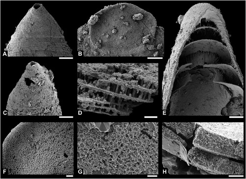
Figure 7. Euconodont elements. A, B, Geniculate coniform element of Drepanodus arcuatus Pander, Citation1856 in lateral and lower view (PMU38390). C, D, Nongeniculate coniform element of Drepanoistodus aff. amoenus (Lindström, Citation1955) sensu Löfgren (Citation1994) in lateral and lower view (PMU38381). E–I, Two elements of Paroistodus numarcuatus (Lindström, Citation1955); nongeniculate coniform element in lateral and lower view (PMU38383) (E, F) and geniculate coniform element in lateral, upper and lower view (PMU38388) (G–I). J–M, Asymmetric nongeniculate coniform (acontiodiform) element of Rossodus aff. manitouensis Repetski & Ethington, Citation1983 in upper (J), posterior (K), lower (L), and anterior (M) view (PMU 38389). N, O, Nongeniculate coniform element of Gen. and sp. indet. in lateral and lower view (PMU38382). Note: Outlines of broken cusps in upper view from respective species illustrated in A, C, E and N. Scale bar equals 100 µm.