Figures & data
Fig. 1. A. Nautilus pompilius. Specimen no. Mo 195106, Solomon Islands. The shell-body attachment band at the apertural margin to show vertical canals that housed epithelial extensions. B, C. Orthoceras regulare. Specimen no. Mo 195101, Kandel. B. Thin, calcified, intermediate sub-layer with vertical pore-canals; the canals are arranged in parallel rows. C. Same specimen in higher magnification. Note the thin calcareous walls of the vertical canals.
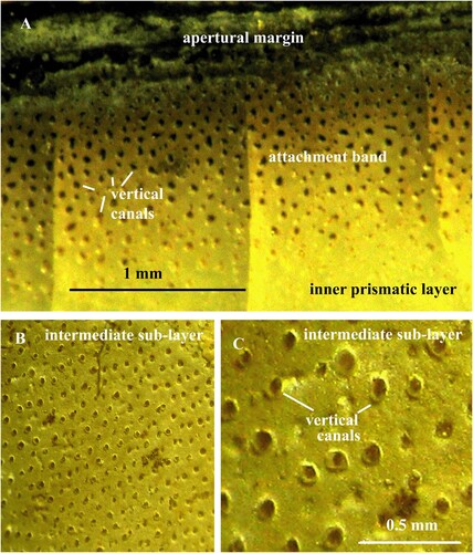
Fig. 2. A. Orthoceras sp. Specimen no. Mo 195102, Runsten. Vertical pore-canals in the intermediate sub-layer and inner prismatic layer; the intermediate sub-layer is rich in organic substance. B. Orthoceras regulare. Same specimen as in B. Thin, calcified, intermediate sub-layer with vertical pore-canals between the nacreous and inner prismatic layers.
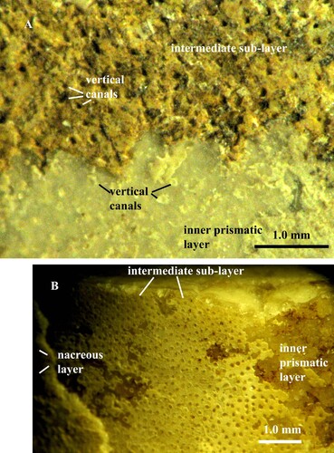
Fig. 3. A, B. Orthoceras scabridum. A, B. Specimen no. Mo 154229, Runsten. A. Intermediate sub-layer with vertical pore-canals B. Canals in higher magnification; note the granular crystalline structure of the intermediate sub-layer
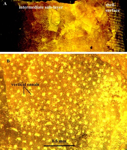
Fig. 4. A, B. Orthoceras sp. Specimen no. Mo 3274, Odensholm. A. Fragments of calcified intermediate sub-layer with numerous vertical pore-canals, arranged in rows. B. Same layer in higher magnification.
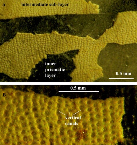
Fig. 5. A, B. Orthoceras sp. Specimen no. Mo 160547, Folkeslunda. A. Intermediate sub-layer with vertical pore-canals. B. Vertical canals in higher magnification; each canal has a wall of globular organic elements. C. Orthoceras sp. Specimen no. Mo 154276, S. Bäck. Intermediate sub-layer with horizontal pore-canals; the canal walls are partially decalcified and consist of globular organic elements; note that the canals are open and empty.
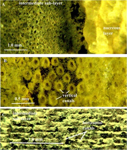
Fig. 6. A, B, C, D, E. Orthoceras sp. Same specimen as in C. Intermediate sub-layer with horizontal pore-canals; note that the globular organic element in the canal walls are connected to each other by strings; the canals are open and empty; in E the vertical canals open into the horizontal canals.
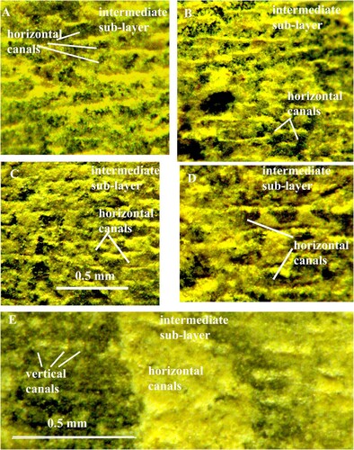
Fig. 7. Orthoceras scabridum. Same specimen as in A. Organic walls of the horizontal pore-canals in higher magnification.
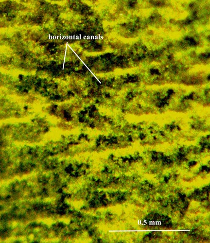
Fig. 8. Orthoceras scabridum. Same specimen as in B. Organic walls of the horizontal pore-canals in higher magnification.
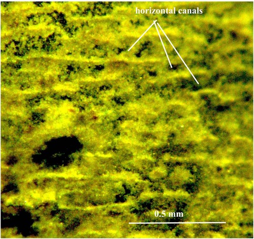
Fig. 9. A, B, C. Orthoceras regulare. Specimen no. Mo 195103, Kandel. A, B. Intermediate sub-layer with horizontal and vertical pore-canals; the vertical canals open into the horizontal canals. C. Intermediate sub-layer with horizontal canals. D. Mitorthoceras perfilosum, Carboniferous orthocerid-like coleoid with horizontal pore-canals (tube-like furrows). Same specimen as in Mutvei & Mapes Citation2019, C. Note the similarity of the horizontal pore-canals in Orthoceras (C) and Mitorthoceras (D).
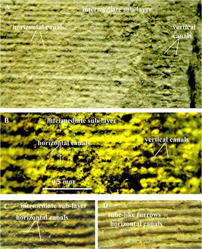
Fig. 10. A, B. Orthoceras regulare. Specimen no. Mo 195104, Kandel. A. Intermediate sub-layer with vertical pore–canals that open into horizontal pore-canals. B. Horizontal pore-canals divided into sections of variable length. C. Orthoceras sp. Specimen no. Mo 195105, Kandel. Intermediate sub-layer with vertical pore-canals; note that the sub-layer consists of organic particles.
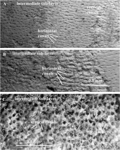
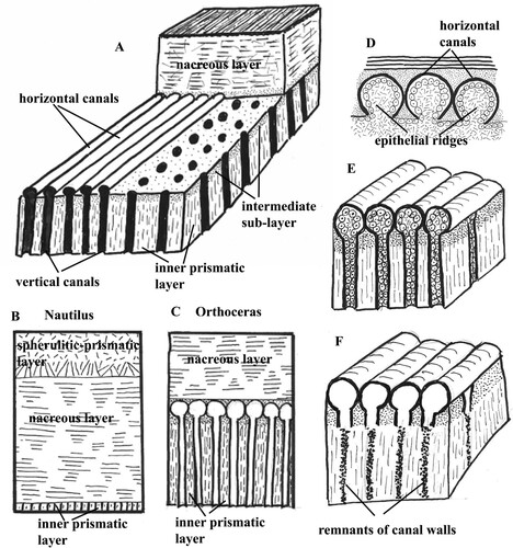
Fig. 11. Schematic presentation of the pore-canals. A. Section of the shell wall in Orthoceras to show arrangement of the horizontal and vertical pore-canals. B. Shell wall in the extant Nautilus. The inner prismatic layer is thin and contains short pores. C. Shell wall in Orthoceras. The inner prismatic layer is thick and contains the pore-canal network that is absent in Nautilus. D. Epithelial ridges on the mantle surface that secreted the walls of the horizontal pore-canals in Orthoceras. E. Horizontal and vertical pore-canals in Orthoceras before diagenesis. F. Horizontal and vertical pore-canals after diagenesis; the walls of the vertical canals are partially dissolved in the inner prismatic layer.