1,053
Views
1
CrossRef citations to date
0
Altmetric
Original Articles

Starfish and Xenopus oocyte maturation

, &
Pages 107-115 | Received 16 Oct 2006, Accepted 05 Dec 2006, Published online: 05 Jun 2007

Figures & data

Figure 1. Scheme showing a starfish oocyte with part of the follicular cells which surround it. A ligand(the gonadostimuline, GS), in the reproductive season stimulates the secretion of the hormone 1‐methyladenine within the follicular cells. A peptide hormone, produced by the radial nerves, elicits 1‐methyladenine excretion from the follicular cells allowing it to bind the oocyte surface where it stimulates a G protein whose βγ subunits cause on one side cAMP decrease and on the other MPF activation with consequent germinal vesicle breakdown. With this breakdown there is further progress toward maturation until metaphase occurs, mediated by MOS and MAPK, but with no activation of the Na+/K+ antiporter, which is activated after oocyte shedding into seawater.
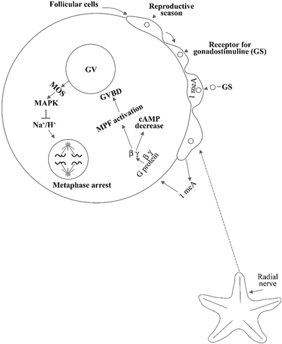
Figure 1. Scheme showing a starfish oocyte with part of the follicular cells which surround it. A ligand(the gonadostimuline, GS), in the reproductive season stimulates the secretion of the hormone 1‐methyladenine within the follicular cells. A peptide hormone, produced by the radial nerves, elicits 1‐methyladenine excretion from the follicular cells allowing it to bind the oocyte surface where it stimulates a G protein whose βγ subunits cause on one side cAMP decrease and on the other MPF activation with consequent germinal vesicle breakdown. With this breakdown there is further progress toward maturation until metaphase occurs, mediated by MOS and MAPK, but with no activation of the Na+/K+ antiporter, which is activated after oocyte shedding into seawater.

Figure 2. (a) Scheme showing the pituitary gonadotropin acting on the follicular cells surrounding the Xenopus oocyte and stimulating progesterone production. Progesterone acts in three ways: one, liberating Cdc25 from the 14‐3‐3 protein and moving it to oocyte pronucleus; second, binding its receptor and therefore inhibiting a G protein from breaking into its subunits; and third, by stimulating binding of the protein CPEB to its site (CPR) in the Mos mRNA. As a consequence of this, Mos mRNA becomes polyadenylated at its 3'end and capped at its 5'end, and therefore translates into its Pp39‐mos protein. This stimulates a MapKK, which in turn activates a MapK and with the aid of Hsp 70 and 90 activates the phosphatase Cdc‐25, which finally activates MPF. Lastly, MPF induces germinal vesicle breakdown. This moves the oocyte to what is described in Figure . (b) Histone phosphorylation which precedes germinal vesicle breakdown makes the oocyte proceed until the II metaphase at which point it becomes arrested again because of the sequential effects of P42, P‐90 RsK, CSF (cytostatic factor), and Emi‐1, until fertilization.
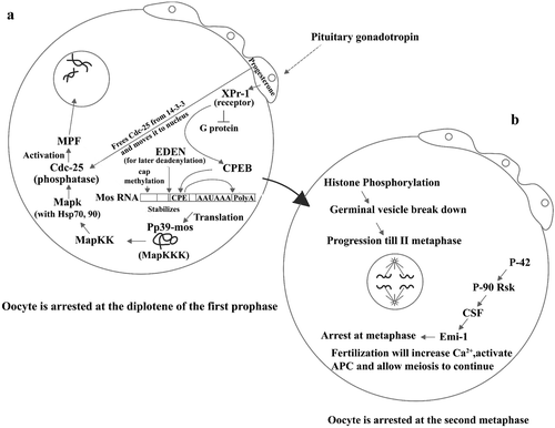
Figure 2. (a) Scheme showing the pituitary gonadotropin acting on the follicular cells surrounding the Xenopus oocyte and stimulating progesterone production. Progesterone acts in three ways: one, liberating Cdc25 from the 14‐3‐3 protein and moving it to oocyte pronucleus; second, binding its receptor and therefore inhibiting a G protein from breaking into its subunits; and third, by stimulating binding of the protein CPEB to its site (CPR) in the Mos mRNA. As a consequence of this, Mos mRNA becomes polyadenylated at its 3'end and capped at its 5'end, and therefore translates into its Pp39‐mos protein. This stimulates a MapKK, which in turn activates a MapK and with the aid of Hsp 70 and 90 activates the phosphatase Cdc‐25, which finally activates MPF. Lastly, MPF induces germinal vesicle breakdown. This moves the oocyte to what is described in Figure 2b. (b) Histone phosphorylation which precedes germinal vesicle breakdown makes the oocyte proceed until the II metaphase at which point it becomes arrested again because of the sequential effects of P42, P‐90 RsK, CSF (cytostatic factor), and Emi‐1, until fertilization.

Figure 3. Comparison between the events leading to the maturation of oocytes of starfish, indicated on the left column, and of amphibians indicated in the right column: in the centre column the events common to the system are reported. The temporal sequence of events is from top to bottom and oblique segments indicate which events precede the other ones.
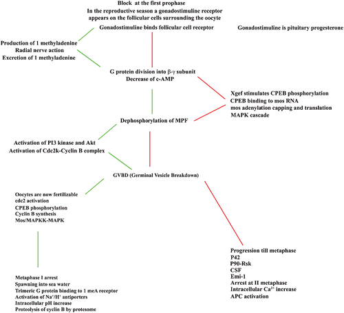
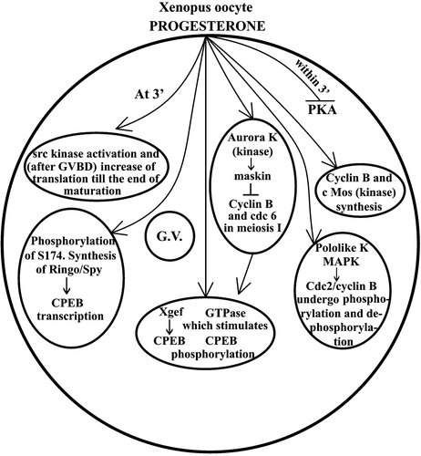
Figure 3. Comparison between the events leading to the maturation of oocytes of starfish, indicated on the left column, and of amphibians indicated in the right column: in the centre column the events common to the system are reported. The temporal sequence of events is from top to bottom and oblique segments indicate which events precede the other ones.

Figure 4. Scheme illustrating recent work on the role of kinase in progesterone‐dependent Xenopus oocyte maturation. The symbol ↑ indicates stimulation, and the symbol ⊥ indicates inhibition. G.V. = germinal vesicle.

Figure 4. Scheme illustrating recent work on the role of kinase in progesterone‐dependent Xenopus oocyte maturation. The symbol ↑ indicates stimulation, and the symbol ⊥ indicates inhibition. G.V. = germinal vesicle.

Reprints and Corporate Permissions

Please note: Selecting permissions does not provide access to the full text of the article, please see our help page How do I view content?

To request a reprint or corporate permissions for this article, please click on the relevant link below:

Academic Permissions

Please note: Selecting permissions does not provide access to the full text of the article, please see our help page How do I view content?

Obtain permissions instantly via Rightslink by clicking on the button below:

If you are unable to obtain permissions via Rightslink, please complete and submit this Permissions form. For more information, please visit our Permissions help page.