452
Views
3
CrossRef citations to date
0
Altmetric
Original Articles

The balancers of Triturus italicus: an ultrastructural approach

, , &
Pages 135-142 | Received 25 Sep 2006, Accepted 08 Dec 2006, Published online: 05 Jun 2007

Figures & data

Figure 1. Triturus italicus embryo and larvae as seen under the binocular microscope. A, embryo at stage 30 of Gallien and Bidaud: the balancer buds (arrow) are conically shaped (bar = 0.5 mm). B, embryo (stage 33 of Gallien and Bidaud) showing a conical–cylindrical balancer (arrow) (bar = 0.5 mm). C, embryo (stage 35 of Gallien and Bidaud); the length of the claviform balancer (arrow) can be compared to that of gill filaments (gf) (bar = 0.2 mm). D, larva (stage 38 of Gallien and Bidaud). After hatching, the balancer (arrow) length increases (gf = gill filaments; bar = 0.1 mm). E, larva (stage 40 of Gallien and Bidaud); the balancers (arrow) reach their largest size (bar = 0.08 mm). F, larva (stage 42 of Gallien and Bidaud); the balancers have completely disappeared (bar = 0.07 mm).
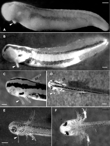
Figure 1. Triturus italicus embryo and larvae as seen under the binocular microscope. A, embryo at stage 30 of Gallien and Bidaud: the balancer buds (arrow) are conically shaped (bar = 0.5 mm). B, embryo (stage 33 of Gallien and Bidaud) showing a conical–cylindrical balancer (arrow) (bar = 0.5 mm). C, embryo (stage 35 of Gallien and Bidaud); the length of the claviform balancer (arrow) can be compared to that of gill filaments (gf) (bar = 0.2 mm). D, larva (stage 38 of Gallien and Bidaud). After hatching, the balancer (arrow) length increases (gf = gill filaments; bar = 0.1 mm). E, larva (stage 40 of Gallien and Bidaud); the balancers (arrow) reach their largest size (bar = 0.08 mm). F, larva (stage 42 of Gallien and Bidaud); the balancers have completely disappeared (bar = 0.07 mm).

Figure 2. Triturus italicus balancers as seen under the SEM. A, balancer at stage 32 of Gallien and Bidaud; the epithelial surface is formed by polygonal pavement cells (bar = 50 µm). B, balancer at stage 38 of Gallien and Bidaud; note the epithelial surface differentiation (PR = proximal region; MR = median region; DR = distal region; bar = 40 µm). C, a higher magnification showing the rather clear margins of pavement cells and the presence of numerous microridges (bar = 5 µm). D, a higher magnification of the median region: the pavement cells appear to be covered by mucus (*) in the central portion of the cells (bar = 4 µm). E, a higher magnification of the apical portion: numerous secretory cones can be recognized (bar = 10 µm). F, secretory cone; each secretory cone appears to have originated from a single pavement cell (bar = 3 µm).
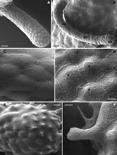
Figure 2. Triturus italicus balancers as seen under the SEM. A, balancer at stage 32 of Gallien and Bidaud; the epithelial surface is formed by polygonal pavement cells (bar = 50 µm). B, balancer at stage 38 of Gallien and Bidaud; note the epithelial surface differentiation (PR = proximal region; MR = median region; DR = distal region; bar = 40 µm). C, a higher magnification showing the rather clear margins of pavement cells and the presence of numerous microridges (bar = 5 µm). D, a higher magnification of the median region: the pavement cells appear to be covered by mucus (*) in the central portion of the cells (bar = 4 µm). E, a higher magnification of the apical portion: numerous secretory cones can be recognized (bar = 10 µm). F, secretory cone; each secretory cone appears to have originated from a single pavement cell (bar = 3 µm).

Figure 3. Triturus italicus balancers: LM and TEM observations. A, light‐microscope micrograph through the subapical portion of the balancers (stage 34 of Gallien and Bidaud); the pavement cells lie on the inner layer of columnar basal cells (PVC = pavement cells; BC = basal cell; bar = 25 µm). B, light‐microscope micrograph through the apical portion of balancers (stage 34 of Gallien and Bidaud); note that the secretory cones (arrow) originate from the central region of the PVCs (* = lipid‐like granules; bar = 25 µm). C, stage 34 of Gallien and Bidaud: a transmission electron micrograph of PVC. Note the numerous elongated or rounded secretion granules (g), which are placed under the apical plasmalemma (bar = 4 µm). D, the ultrastructural features of PVC in the apical region of a well‐developed balancer (stage 38 of Gallien and Bidaud). The whole cytoplasm is filled by granules (g) and bundles of tonofilaments (arrowheads) (bar = 2 µm). E, a higher magnification showing the microridges embedded in the fuzzy coat of mucus (*). The secretory granules (g) are arranged in 5–7 layers (stage 38 of Gallien and Bidaud; bar = 500 nm). F, well‐developed rough endoplasmic reticulum (RER) of the inner layer cells (stage 38 of Gallien and Bidaud; n = nucleus; bar = 1 µm).
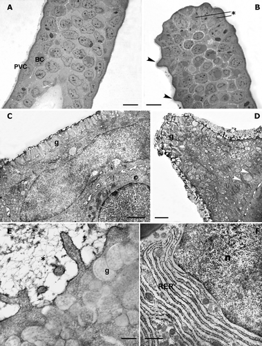
Figure 3. Triturus italicus balancers: LM and TEM observations. A, light‐microscope micrograph through the subapical portion of the balancers (stage 34 of Gallien and Bidaud); the pavement cells lie on the inner layer of columnar basal cells (PVC = pavement cells; BC = basal cell; bar = 25 µm). B, light‐microscope micrograph through the apical portion of balancers (stage 34 of Gallien and Bidaud); note that the secretory cones (arrow) originate from the central region of the PVCs (* = lipid‐like granules; bar = 25 µm). C, stage 34 of Gallien and Bidaud: a transmission electron micrograph of PVC. Note the numerous elongated or rounded secretion granules (g), which are placed under the apical plasmalemma (bar = 4 µm). D, the ultrastructural features of PVC in the apical region of a well‐developed balancer (stage 38 of Gallien and Bidaud). The whole cytoplasm is filled by granules (g) and bundles of tonofilaments (arrowheads) (bar = 2 µm). E, a higher magnification showing the microridges embedded in the fuzzy coat of mucus (*). The secretory granules (g) are arranged in 5–7 layers (stage 38 of Gallien and Bidaud; bar = 500 nm). F, well‐developed rough endoplasmic reticulum (RER) of the inner layer cells (stage 38 of Gallien and Bidaud; n = nucleus; bar = 1 µm).

Figure 4. Triturus italicus balancers: TEM observations. A, Golgi apparatus (G) surface in the inner epithelial layer cell (stage 38 of Gallien and Bidaud; bar = 200 nm). B, a large lipid‐like granule (lg) is placed near the nuclear surface in the inner epithelial layer cell; n = nucleus; (stage 38 of Gallien and Bidaud; bar = 1 µm). C, numerous bundles of tonofilaments (*) can be observed in the epithelial cells: their amount increases as the development proceeds (stage 38 of Gallien and Bidaud; bar = 330 nm). D, a transmission electron micrograph of unmyelinated neuritis present among the epithelial cells (stage 38 of Gallien and Bidaud; bar = 1µm). E, a transmission electron micrograph of neurites, enveloped by a Schwann cell, in the basal layer of a well‐innervated epithelium (stage 38 of Gallien and Bidaud; bar = 330 nm). F, a transmission electron micrograph of a degenerating pavement cell in the balancers of a Triturus italicus larva at stage 40 of Gallien and Bidaud. Note the large cytoplasmic lacunae (*) (stage 40 of Gallien and Bidaud; bar = 2 µm).
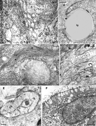
Figure 4. Triturus italicus balancers: TEM observations. A, Golgi apparatus (G) surface in the inner epithelial layer cell (stage 38 of Gallien and Bidaud; bar = 200 nm). B, a large lipid‐like granule (lg) is placed near the nuclear surface in the inner epithelial layer cell; n = nucleus; (stage 38 of Gallien and Bidaud; bar = 1 µm). C, numerous bundles of tonofilaments (*) can be observed in the epithelial cells: their amount increases as the development proceeds (stage 38 of Gallien and Bidaud; bar = 330 nm). D, a transmission electron micrograph of unmyelinated neuritis present among the epithelial cells (stage 38 of Gallien and Bidaud; bar = 1µm). E, a transmission electron micrograph of neurites, enveloped by a Schwann cell, in the basal layer of a well‐innervated epithelium (stage 38 of Gallien and Bidaud; bar = 330 nm). F, a transmission electron micrograph of a degenerating pavement cell in the balancers of a Triturus italicus larva at stage 40 of Gallien and Bidaud. Note the large cytoplasmic lacunae (*) (stage 40 of Gallien and Bidaud; bar = 2 µm).

Reprints and Corporate Permissions

Please note: Selecting permissions does not provide access to the full text of the article, please see our help page How do I view content?

To request a reprint or corporate permissions for this article, please click on the relevant link below:

Academic Permissions

Please note: Selecting permissions does not provide access to the full text of the article, please see our help page How do I view content?

Obtain permissions instantly via Rightslink by clicking on the button below:

If you are unable to obtain permissions via Rightslink, please complete and submit this Permissions form. For more information, please visit our Permissions help page.