540
Views
5
CrossRef citations to date
0
Altmetric
Original Articles

Some steps of spermatogenesis in Halichondria semitubulosa (Demospongiae, Halichondriidae)

, , , &
Pages 117-122 | Received 05 Oct 2006, Accepted 21 Dec 2006, Published online: 05 Jun 2007

Figures & data

Figure 1. Halichondria semitubulosa. A, sponge specimens in their natural environment (scale bar, 2 cm); B, semithin section showing choanocytes chambers (ChC) very close to one another (scale bar, 12 µm); C, Semithin section showing a spermatocyst (SC) near a choanocytes chamber (ChC) (scale bar, 12 µm); D, ultrathin section showing a spermatocyst (SC) and a choanocytes chamber (ChC). Note the debris (De) included in the cytoplasmic matrix. Sp I, spermatocytes of the first order (scale bar, 2 µm).
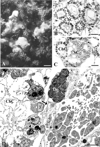
Figure 1. Halichondria semitubulosa. A, sponge specimens in their natural environment (scale bar, 2 cm); B, semithin section showing choanocytes chambers (ChC) very close to one another (scale bar, 12 µm); C, Semithin section showing a spermatocyst (SC) near a choanocytes chamber (ChC) (scale bar, 12 µm); D, ultrathin section showing a spermatocyst (SC) and a choanocytes chamber (ChC). Note the debris (De) included in the cytoplasmic matrix. Sp I, spermatocytes of the first order (scale bar, 2 µm).

Figure 2. TEM micrographs of spermatogenetic phases.A, two spermatocysts (SC) at different stage of maturation. Note the layer of pinacocyte‐like cells (P) delimiting the cysts. F, flagella (scale bar, 5 μm); B, Spermatocytes of the first order (Sp I). M, mitochondria; N, nucleus (scale bar, 0.7 µm); C, spermatocytes of the second order (Sp II) connected by a cytoplasmic bridge (arrow). M, mitochondrion; N, nucleus (scale bar, 0.5 µm); D, a large mitochondrion (M) in the spermatocyte of the second order. Gl, glycogen; I, round‐shaped electron‐dense inclusions; N, nucleus (scale bar, 0.2 µm); E, spermatid. Note the presence of glycogen in the basal region of the tail (arrows). Gl, glycogen; I, electron‐dense inclusions; M, mitochondrion; N, nucleus (scale bar, 0.4 µm).
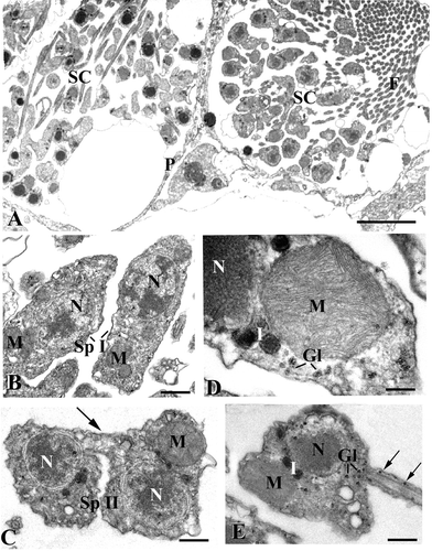
Figure 2. TEM micrographs of spermatogenetic phases.A, two spermatocysts (SC) at different stage of maturation. Note the layer of pinacocyte‐like cells (P) delimiting the cysts. F, flagella (scale bar, 5 μm); B, Spermatocytes of the first order (Sp I). M, mitochondria; N, nucleus (scale bar, 0.7 µm); C, spermatocytes of the second order (Sp II) connected by a cytoplasmic bridge (arrow). M, mitochondrion; N, nucleus (scale bar, 0.5 µm); D, a large mitochondrion (M) in the spermatocyte of the second order. Gl, glycogen; I, round‐shaped electron‐dense inclusions; N, nucleus (scale bar, 0.2 µm); E, spermatid. Note the presence of glycogen in the basal region of the tail (arrows). Gl, glycogen; I, electron‐dense inclusions; M, mitochondrion; N, nucleus (scale bar, 0.4 µm).

Figure 3. TEM micrographs.A, spermatozoon with electron‐dense nucleus (N) and inclusions (I) (scale bar, 0.5 µm); B, section of a spermatozoon showing the co‐presence of nucleus (N) and mitochondrion (M). I, inclusions (scale bar, 0.4 µm); C, an almost empty cyst including only some spermatozoa (S) (scale bar, 6 µm).
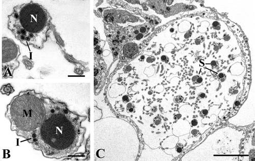
Figure 3. TEM micrographs.A, spermatozoon with electron‐dense nucleus (N) and inclusions (I) (scale bar, 0.5 µm); B, section of a spermatozoon showing the co‐presence of nucleus (N) and mitochondrion (M). I, inclusions (scale bar, 0.4 µm); C, an almost empty cyst including only some spermatozoa (S) (scale bar, 6 µm).

Reprints and Corporate Permissions

Please note: Selecting permissions does not provide access to the full text of the article, please see our help page How do I view content?

To request a reprint or corporate permissions for this article, please click on the relevant link below:

Academic Permissions

Please note: Selecting permissions does not provide access to the full text of the article, please see our help page How do I view content?

Obtain permissions instantly via Rightslink by clicking on the button below:

If you are unable to obtain permissions via Rightslink, please complete and submit this Permissions form. For more information, please visit our Permissions help page.