Figures & data
Table I. Mean total lengths (mm), body and gonadal mass (g) of the female chub Leuciscus cephalus by collection.
Table II. Maturity stages of ovaries of wild‐caught chub Leuciscus cephalus. Oocyte stage, appearance and oocyte diameter (mean±SD) detected monthly.
Figure 1 Histological sections(A–D) of ovaries of the chub Leuciscus cephalus during A, undeveloped stage: immature (primitive oogonia, PO); B, early recovery of gonadal activity stage: maturing (early vitellogenic, EV); C, ripening stage: mature (vitellogenic, VO); D, resting stage: post‐mature (atretic, AT). Stained with hematoxylin and eosin. Scale bars: A–D = 50 µm and A'–D' = 100 µm.
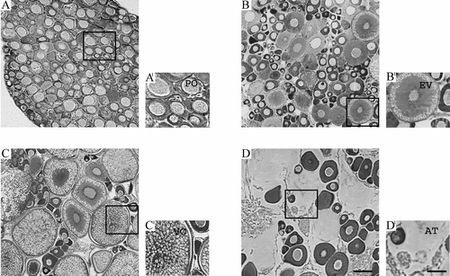
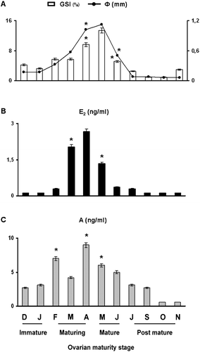
Figure 2 Monthly changes ofφ = mean oocyte diameter and GSI = gonadosomatic index (A), plasma levels of E2 = 17β‐estradiol (B); and A = androgen (C) in the chub Leuciscus cephalus. Each value represents the mean of three determinations in triplicate of each sample (n = 20)±SD. * Level of significance versus the mean values observed in the preceding month (P<0.05).