626
Views
8
CrossRef citations to date
0
Altmetric
COMPARATIVE MORPHOLOGY & PHYSIOLOGY

Immunohistochemical analysis of the adhesive papillae of Botrylloides leachi (Chordata, Tunicata, Ascidiacea): Implications for their sensory function

, , &
Pages 325-329 | Received 21 Dec 2006, Accepted 19 Mar 2007, Published online: 30 Nov 2007

Figures & data

Figure 1 A, trunk region of a Botrylloides leachi larva, showing the disposition of the left dorsal and the ventral papilla. The right dorsal one is not focused. B, longitudinal histological section of the trunk region showing the wide hemocoelic region under the anterior ectoderm. C,D, two sections at different level of a dorsal right papilla. E, histological section of the anterior ectoderm, showing the cluster of secreting cells protruding from the tunic. F,G, histological sections at different level of the cluster of secreting cells. bc, blood cells, cn, central neurons; dp, dorsal papilla; ilt, inner layer of the tunic; l, lacuna; ph, photolith; olt ,outer layer of the tunic; pn, peripheral neurons; sc, secreting cells, vp, ventral papilla. Scale bar: A,B 100 µm; C–G 20 µm.
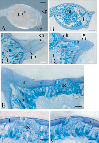
Figure 1 A, trunk region of a Botrylloides leachi larva, showing the disposition of the left dorsal and the ventral papilla. The right dorsal one is not focused. B, longitudinal histological section of the trunk region showing the wide hemocoelic region under the anterior ectoderm. C,D, two sections at different level of a dorsal right papilla. E, histological section of the anterior ectoderm, showing the cluster of secreting cells protruding from the tunic. F,G, histological sections at different level of the cluster of secreting cells. bc, blood cells, cn, central neurons; dp, dorsal papilla; ilt, inner layer of the tunic; l, lacuna; ph, photolith; olt ,outer layer of the tunic; pn, peripheral neurons; sc, secreting cells, vp, ventral papilla. Scale bar: A,B 100 µm; C–G 20 µm.

Figure 2 Confocal laser images of dorsal right(A) and left (B) papillae showing the localization of serotonin (FITC signal, in green) and of β‐tubulin (TRITC signal, in red). The yellow colour at the apex is an artifact due to unspecific adhesion of both secondary antibodies in the fenestration of the tunic, where distal processes of central neurons reach the surface. C,D, superimposition of A and B to light microscopy imagines. cn, central neurons; ilt, inner layer of the tunic; pn, peripheral neurons. Scale bar: 20 µm.
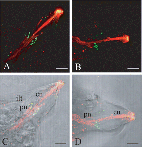
Figure 2 Confocal laser images of dorsal right(A) and left (B) papillae showing the localization of serotonin (FITC signal, in green) and of β‐tubulin (TRITC signal, in red). The yellow colour at the apex is an artifact due to unspecific adhesion of both secondary antibodies in the fenestration of the tunic, where distal processes of central neurons reach the surface. C,D, superimposition of A and B to light microscopy imagines. cn, central neurons; ilt, inner layer of the tunic; pn, peripheral neurons. Scale bar: 20 µm.
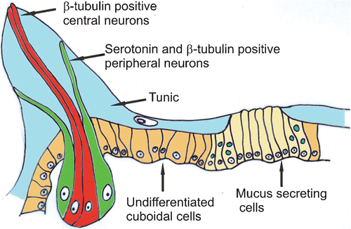
Figure 3 Schematic drawing of the anterior ectoderm reconstructed comparing histological sections and confocal laser images.

Figure 3 Schematic drawing of the anterior ectoderm reconstructed comparing histological sections and confocal laser images.

Reprints and Corporate Permissions

Please note: Selecting permissions does not provide access to the full text of the article, please see our help page How do I view content?

To request a reprint or corporate permissions for this article, please click on the relevant link below:

Academic Permissions

Please note: Selecting permissions does not provide access to the full text of the article, please see our help page How do I view content?

Obtain permissions instantly via Rightslink by clicking on the button below:

If you are unable to obtain permissions via Rightslink, please complete and submit this Permissions form. For more information, please visit our Permissions help page.