Figures & data
Figure 1 Map of localities and occurrences of Pseudocirrhipathes mapia in the Marine Park of Bunaken. 1, Mapia (type locality) (Coast of Manado); 2, Onong (Siladen Island); 3, Likuan 3 (Bunaken Island); 4, Nain (Nain Island); 5, Wreck of Manado (Coast of Manado); 6, Raymond's Point (Manado Tua Island).
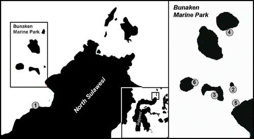
Figure 2 Comparison of the contracted polyps of the three unbranched genera. A, Cirrhipathes; B, Stichopathes; C, Pseudocirrhipathes.
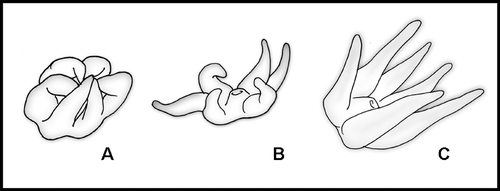
Figure 3 Spines of Pseudocirrhipathes mapia. A, spiral arrangement of spines along the basal portion of the stem; B, close up view of the basal region; C,D, slightly tuberculated triangular spines in the basal region; E, close up view of the central region, where spines are arranged in regular longitudinal rows; F, tuberculated sub‐triangular spines in the intermediate region; G,H, circular arrangement of tubercules on the surface of spines situated in the central portions of the colony; I,J, different arrangement of tubercules on the proximal side of spines, either covering the entire surface or absent. The tip of the spines is always free of tubercules; K, close‐up view of the apical region, where the spines are arranged into well‐separated verticils. Ridges were produced by the collapse of the skeleton due to dehydration; L, smooth triangular spines in the apical verticils; M,N, variation in diameter of the central canal in the central and apical portion of the colony, respectively. Scale bars: A, 1 mm. B, E, 0.5 mm. K, M, N, 0.2 mm. C, D, F–J, L, 0.1 mm.
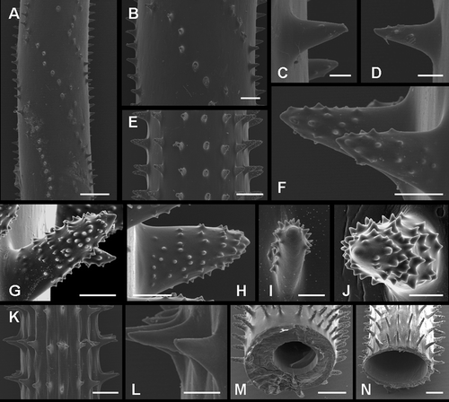
Figure 4 Underwater photographs of Pseudocirrhipathes mapia. A, macro‐photograph of P. mapia showing an expanded polyp surrounded by resting zooids that do not completely contract their tentacles; B, abpolypar side of a P. mapia colony showing the polyp‐free side of the axis; C, polypar side of a P. mapia colony; D, macro‐photograph of P. mapia showing expanded polyps. Sagittal tentacles are inserted at a lower level; E, two colonies of P. mapia; F, expanded polyp of P. mapia showing the double margin mouth; G, open mouth of an egesting polyp; H, a large discharged basitrich isorhiza, showing the spiny stylet and the incompletely extruded filament; I, patch distribution of nematocysts on the ectodermal surface of the tentacle; J, colony of P. mapia hiding a mimicking Pontonides unciger; K, extremely long sweeper tentacles of a white P. mapia, touching a dead Acropora. Images A, D, F, G, J are courtesy of Massimo Boyer. Scale bars: H, 25 µm. G, I, 1 mm. A, D, F, J, 2 mm. B, C, 5 mm. K, 20 mm. E, 20 cm.
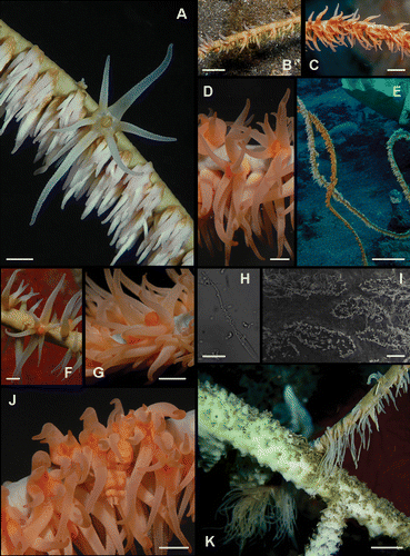
Figure 5 Spines of Cirripathes rumphii. Coel. 02559b A, arrangement of spines in the apical portion of the colony; B, close up view of a verticil of spines; C,D, highly tuberculated spines; E, diameter of the central canal. Coel. 02559a F, arrangement of spines in the apical portion of the colony; G,H, close‐up views of the apically tuberculated spines; I, frontal view of the laterally compressed spines. Scale bars: C, D, 50 µm. B, G, H, 100 µm. I, 200 µm. A, E, F, 500 µm.
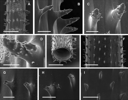
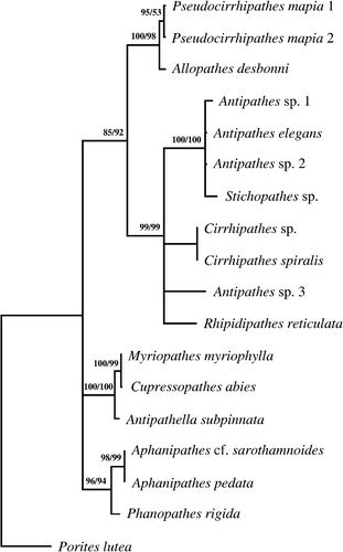
Figure 6 Phylogenetic tree. Consensus tree obtained by maximum parsimony (length 673; CI 0.792; RI 0.793) and maximum likelihood for 16 antipatharian species based on alignment (1.030 positions long and containing 206 parsimony informative sites) of internal transcribed spacer rDNA sequences. Numbers along the branches represent bootstrap estimates: MP value (left) and ML value (right).