Figures & data
Figure 1. Retinal section of 10-day chick embryos. A, H&E-stained section showing different layers of retina; B, section showing ill defined layering of retina (H&E stain); C, Toludine blue-stained high-resolution section. The arrow is pointing to a nucleus undergoing mitosis. The arrowhead is pointing external limiting membrane, while rod-shaped pigment granules are seen in the pigment epithelium; D, Toludine blue-stained high-resolution section showing inner nuclear layer mostly having elongated nuclei. The bar shown at the bottom of the figure represents approximately in A, 50 μm. B, 65 μm. C, 15 μm. D, 10 μm.
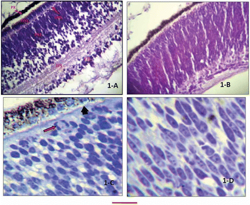
Table I. Comparison of chicken retinal layers thickness at different developmental stages
Table II. Comparison of nuclear shapes of inner and outer nuclear cell layers at different developmental stages of chicken retinae
Figure 2. Retinal differentiation stages. A, retinal section of 15-day chick embryo (H&E stain), showing all layers, starting from top, pigment epithelium, rods and cones (abutting tightly into the epithelium, compromising the measureable height/thickness of this layer), vague impression of external limiting membrane, ONL, a thin OPL, well-developed INL, IPL, ganglion cell layer and internal limiting membrane; B, retinal section of a newly hatched chick (H&E stain) showing morphological differences from day 15 retina, with obviously elongated photoreceptor cells protruding in the pigment epithelium producing vacuolated appearances. The INL is compressed between the tall photoreceptor cells and markedly developed IPL; C, magnified view of INL of 15-day embryo (H&E stain), showing interconnections between the inner nuclear and ganglion cell layer; D, retinal section of 15-day chick embryo (Cresyl violet stain). The pale-looking external limiting layer and external plexiform layers (not very clear in A) are highlighted. A few ganglion cells are seen migrating down the inner plexiform layer. The bar shown at the bottom of the figure represents approximately in A, B, D, 40 μm. C, 15 μm.
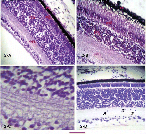
Figure 3. High-resolution, Toludine Blue-stained day 15 retinal sections. A, the section shows rod-shaped pigment granules and mostly oval nuclear morphology of the outer nuclear layer; B, the arrow marks the external limiting membrane stretching through the outer and inner segments of the photoreceptor cells. A few bipolar nuclei are identified in the inner nuclear layer (arrowheads). The bar shown at the bottom of the figure represents approximately in A, 10 μm. B, 15 μm.
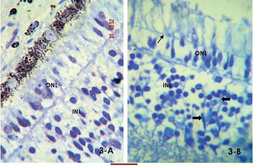
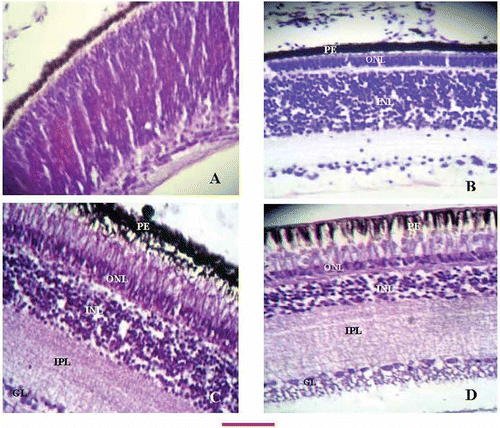
Figure 4. Retinal sections at different developmental ages of chicken. A, 10-day embryo retinal section showing undifferentiated layers. Appearance of a thin, inner plexiform layer is just demarcating the nuclear and ganglion cell layers; B, identifiable external limiting layer is stretching underneath the epithelium dividing the upper and lower segments of the photoreceptor cells. The OPL is present between the outer and inner nuclear layers. The thickness of the INL can be appreciated to be more as compared to the more advanced ages (C and D); C, at the time of hatching the sections revealed marked elongation of the phoreceptor cells, mature morphology of the ONL, identifiable OPL, a compact INL and broad band of the OPL; D, retinal sections of an adult chicken showing prominent vacuolation of the epithelium, thin outer nuclear layer, a more compromised ONL as compared to B and C, a very broad INL, and a single layer of ganglion cells (compare with A and B). The bar shown at the bottom of the figure represents approximately A, B, 60 μm. C, D, 50 μm.