Figures & data
Figure 1. Hemerobiidae. Wesmaelius subnebulosus. A, Interference-contrast micrograph of the anterior region of a living sperm. Note the two thin prolongations; B, Longitudinally sectioned spermatozoa showing a bilayered acrosome (a), the nucleus (N) and the two prolongations of the apical region (arrowheads); C, Cross-sectioned sperm at the level of the nuclei (N). The condensed nuclear material is surrounded by a nuclear envelope that fans out laterally into two differently long thin wings (arrows). Note the dense material (asterisks) surrounding the sperm cells; D, Cross-section through testes showing early spermatids. At this level a layer of microtubules (mt) surrounds the nucleus which shows dense chromatin filaments; E, Cross-section through the posterior region of spermatids showing the nuclei (N) with a triangular shape; mt, microtubules; F, Cross-section through the anterior region of spermatids with the lenticular nuclei (N); m, mitochondria; ax, axoneme; G, Cross-section through an early spermatid showing the basal body provided with microtubule triplets and two mitochondrial derivatives (m).
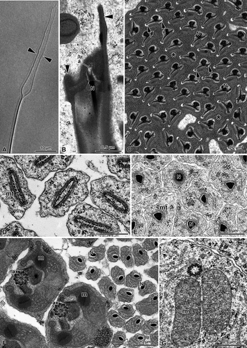
Figure 2. Hemerobiidae. Wesmaelius subnebulosus. A,B, Cross-sections through several sperm tails, each showing a 9+9+2 axoneme (ax), the accessory bodies (ab) and two mitochondrial derivatives (m). Note the basal body region (bb) consisting of a bundle of microtubular elements embedded in a dense material; arrows indicate the plasma membrane grooves. N, nucleus.
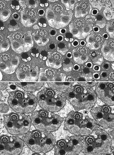
Figure 3. Hemerobiidae. A, Cross-sections through the sperm tails of Wesmaelius subnebulosus. Two mitochondrial derivatives (m) flank the axoneme (ax). Note that in the region close to the axoneme each mitochondrial derivative contains crystalline material (asterisks). This material shows a dense mid ridge (arrowheads). The centriolar region (c) consists of a bundle of microtubular elements embedded in a dense material; B, A membrane specialization is visible at the level of a groove beneath the plasma membrane (arrowheads). Note that the centriole adjunct material (ca) surrounds the axoneme (ax). The flagellum shows a 9+9+2 axonemal pattern with accessory tubules (at) provided with 16 protofilaments. Note that each mitochondrial derivative (m) has two finger-like projections and a crystallized material in the matrix (asterisk); C, Cross-section through the most posterior end of the sperm tail which contains only the axoneme (ax); D–G, Spermatozoa of Hemerobius micans cross-sectioned at the level of the nuclei (N). Note that they are embedded in a dense material and consist of a thin axial region and two long lateral wings (arrows); H, Cross-section through the sperm tails of H. micans showing a 9+9+2 axoneme (ax), two elongated accessory bodies (ab) and the two mitochondrial derivatives (m). Note the asymmetrical shape of the sperm tail.
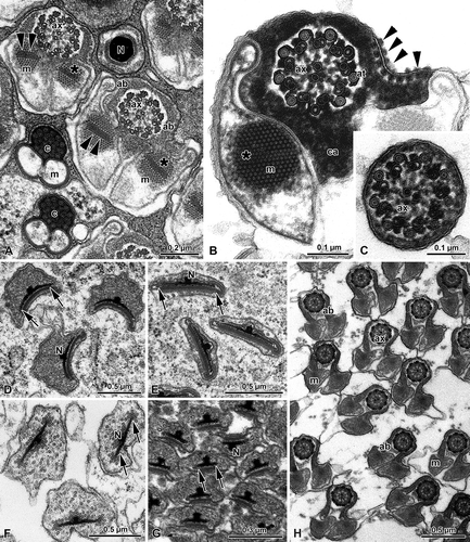
Figure 4. Chrysopidae. A, Spermatozoa of Dichocrysa prasina and B, Chrysopa formosa cross-sectioned at the level of the nuclei (N). The cylindrical nucleus (N) shows the nuclear envelope that expands in a thin wing (arrows). The nucleus is surrounded by a great amount of dense paracrystalline material (p) and by a bundle of parallel rodlets (r); C,E, Cross-sectioned sperm tails of D. prasina and D, Chrysopa intima showing the two mitochondrial derivatives (m) that surround most of the axoneme (ax) and leave only a little space for the accessory bodies (ab). The flagellum shows a 9+9+2 axoneme. Note the bridges (arrowheads) connecting the axonemal doublets 2 and 5 to the mitochondrial derivatives.
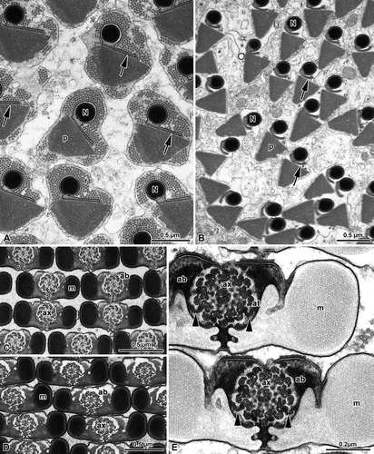
Figure 5. Mantispidae. Perlamantispa perla. A, Cross-sectioned sperm at the level of the nuclei (N). Each nucleus consists of a dense cylindrical axial part which expands laterally into two differently long thin wings (arrows). The nucleus is surrounded by a dense material; B, Cross-sectioned euspermatozoon showing long accessory bodies (ab) partially flanking the axoneme (ax). The flagellum has a 9+9+2 axoneme with accessory tubules (at) provided with 16 protofilaments in their tubular wall. Note the beak-like shape of the intertubular material (im) adhering to the accessory tubules; m, mitochondrial derivatives.
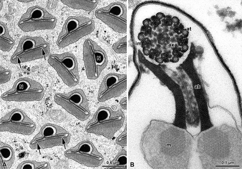
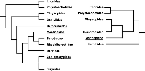
Figure 6. Phylogenetic relationships based on holomorphological and molecular characters. The latter data are also supported by the spermatological results reported in the paper (from Haring & Aspöck Citation2004).