Figures & data
Figure 1. Platycleis intermedia, cross-section of seminal receptacle. c, cuticle; e, epithelium; L, lumen of seminal receptacle; mu, muscle. Haematoxylin–Eosin. Scale bar: 50 mm.
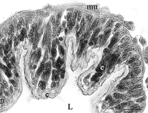
Figure 2. Diagram of the ultrastructural organization of the epithelium of the seminal receptacle and spermathecal duct of Platycleis intermedia. 1, cuticle-forming cell; 2, gland cell. bl, basal lamina; ci, cuticular intima; g, Golgi bodies; gr, electron-dense granules; m, mitochondria; mt, microtubules; n, nucleus; r, reservoir; RER, rough ergastoplasmic reticulum; v, vesicle.
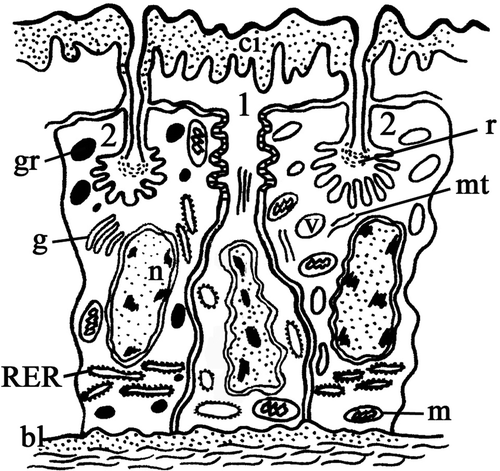
Figure 3. Platycleis intermedia, seminal receptacle. A, the cuticle-forming cells (cc) and the gland cells (gc) are tightly packed. The perinuclear cistern appears dilated (arrow); RER, rough ergastoplasmic reticulum; n, heterochromatic nucleus; v, vesicles. B, the contiguous membranes form deep interdigitation with septate junctions (arrows); C, the microtubules (mt) in the cuticle-forming cells; D, the cuticle-forming cells around the apical region form sac-like dilations (little square). Inset: detail of the dilations in the little square; c, cuticle; gc, gland cell; gr, electron-dense granules; m, mitochondria; mi, microvilli of the end-apparatus; n, nucleus. E, apical region. Arrow: osmiophilic layer of epicuticle; end, endocuticle; ep, granular layer of epicuticle. F, gland cell. RER, rough ergastoplasmic reticulum is mainly made up of flattened and parallel cisterns. Scale bars: A, 92 nm. B, 1920 nm. C, 140 nm. D, 3390 nm. E, 480 nm. F, 870 nm.
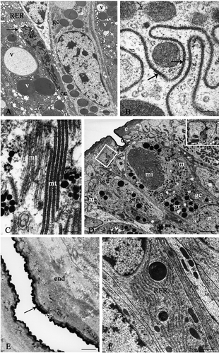
Figure 4. Platycleis intermedia. A and B, seminal receptacle. A, the wall of the efferent duct consists solely of an epicuticle (ep) that is thinner at the reservoir (headarrow) and immediately after thickens and remains constant throughout its course (arrow); fw, felt-work. B, basal portion. Arrow, sediment of basal lamina; bl, basal lamina; e, epithelium; mu, muscular layer. C and D, connecting tract of the spermathecal duct: cuticle-forming cells. C, end, endocuticle; m, mitochondria; s, septate junctions. D, ae, apical expansions; v, vesicles with heterogeneous content. Scale bars: A, 650 nm. B, 1920 nm. C, 870 nm. D, 1510 nm.
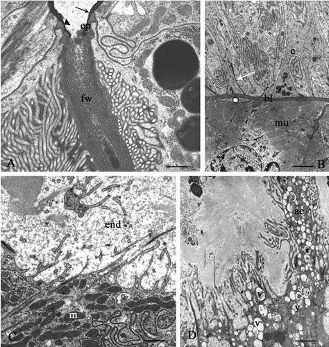
Figure 5. A and B: Platycleis intermedia spermathecal duct. A, proximal tract, apical region of the cuticle-forming cells. Arrows, points of attachment of microtubules; end, endocuticle; mi, microvilli. B, basal region. Arrows, infoldings of the plasma membrane; bl, basal lamina; m, mitochondria. C–E: Platycleis grisea, seminal receptacle. C, gland cell. v, vesicles with heterogeneous content; D, vesiscles (v) with low electrondense content near to central reservoir (r); E, ed, efferent duct; v, vesicles with heterogeneous content. Scale bars: A, 1120 nm. B, 1920 nm. C, 870 nm. D, 870 nm. E, 650 nm.
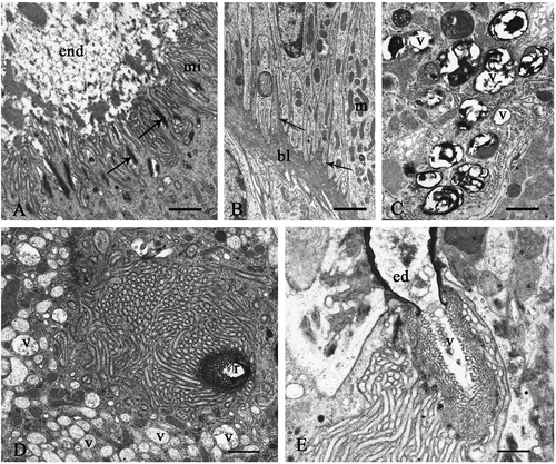
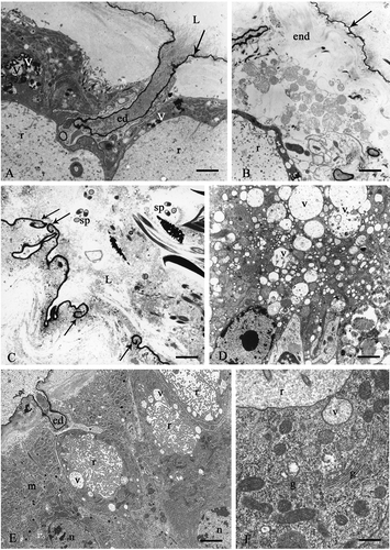
Figure 6. Sepiana sepium. A, seminal receptacle. Arrow, cuticle; ed, efferent duct; L, lumen; r, reservoir; v, vesiscles with heterogeneous content. B, proximal tract of the spermathecal duct. Cuticle-forming cells. Arrow, osmiophilic layer of the epicuticle; end, endocuticle; r, reservoir. C, arrows, break of the osmiophilic layer of the epicuticle; L, lumen; sp, sperms. D, the cytoplasm of the gland cells has numerous vesicles (v) with a low electron-dense content. E and F Tessellana tessellata, seminal receptacle. E, vesicles (v) in the cytoplasm around the reservoir (r); ed, efferent duct; m, mitochondria; n, nucleus. F, gland cells. g, Golgi bodies; r, reservoir; v, vesicle with a granular content opening into the reservoir. Scale bars: A, 1510 nm. B, 1920 nm. C, 1120 nm. D, 1920 nm. E, 2000 nm. F, 480 nm.