1,066
Views
8
CrossRef citations to date
0
Altmetric
Original Articles

The rete mirabile of the tail, an effective site for sampling sterile blood from armadillos (Dasypodidae, Xenarthra)

, , &
Pages 63-69 | Received 29 Mar 2010, Accepted 31 Jul 2010, Published online: 07 Mar 2011

Figures & data

Table I. Number of specimens used for anatomo-histological studies, field blood collection and lymphocyte culture

Figure 1. a, Restraint in Zaedyus pichiy; b, blood collection in C. villosus from the tail between the first and the second rings. Scale bar: 3 cm.
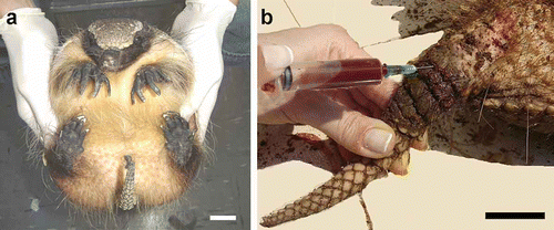
Figure 1. a, Restraint in Zaedyus pichiy; b, blood collection in C. villosus from the tail between the first and the second rings. Scale bar: 3 cm.

Figure 2. a, Individual of C. vellerosus in immobilization device; b, blood collection by a single operator using immobilization device; c, schematic drawing of immobilization device. I: Polyvinyl chloride pipe, II: trigger, III: wooden base, IV: rod, V: iron plate lined with EVA foam. Scale bar: 3 cm.

Figure 2. a, Individual of C. vellerosus in immobilization device; b, blood collection by a single operator using immobilization device; c, schematic drawing of immobilization device. I: Polyvinyl chloride pipe, II: trigger, III: wooden base, IV: rod, V: iron plate lined with EVA foam. Scale bar: 3 cm.

Figure 3. Tail transverse section of Chaetophractus villosus. HE staining. Scale bar: 2.5 mm. RM: rete mirabile of the medial coccygeal artery. MAR: main artery of the rete mirabile, cv: coccygeal vertebrae, bdp: bony dermal plate, mvs: medial ventral sacrocaudal muscle, lvs: lateral ventral sacrocaudal muscle, cvi: caudal ventral intertransverse muscle, cdi: caudal dorsal intertransverse muscle, lds: lateral dorsal sacrocaudal muscle, mds: medial dorsal sacrocaudal muscle, hf: hair follicle.
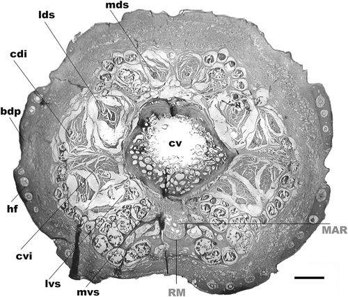
Figure 3. Tail transverse section of Chaetophractus villosus. HE staining. Scale bar: 2.5 mm. RM: rete mirabile of the medial coccygeal artery. MAR: main artery of the rete mirabile, cv: coccygeal vertebrae, bdp: bony dermal plate, mvs: medial ventral sacrocaudal muscle, lvs: lateral ventral sacrocaudal muscle, cvi: caudal ventral intertransverse muscle, cdi: caudal dorsal intertransverse muscle, lds: lateral dorsal sacrocaudal muscle, mds: medial dorsal sacrocaudal muscle, hf: hair follicle.

Figure 4. Tail transverse sections of the following armadillos. a, Dasypus hybridus; b, Chaetophractus vellerosus; c, Tolypeutes matacus; d, Euphractus sexcinctus. Scale bar: 1 mm. Inset histological images of the rete mirabile. HE staining. Scale bar: 200 µm. RM: rete mirabile of the medial coccygeal artery, MAR: main artery of the rete mirabile, LCV: lateral caudal veins, cv: coccygeal vertebrae, sp: spinous process, tp: transverse process, bdp: bony dermal plate, mvs: medial ventral sacrocaudal muscle, lvs: lateral ventral sacrocaudal muscle, cvi: caudal ventral intertransverse muscle, cdi: caudal dorsal intertransverse muscle, lds: lateral dorsal sacrocaudal muscle, mds: medial dorsal sacrocaudal muscle.
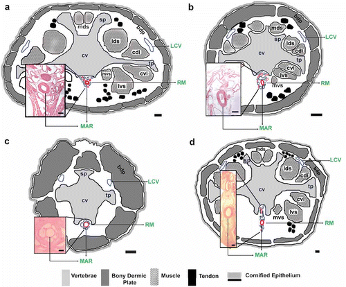
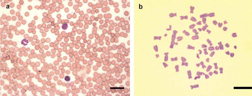
Figure 4. Tail transverse sections of the following armadillos. a, Dasypus hybridus; b, Chaetophractus vellerosus; c, Tolypeutes matacus; d, Euphractus sexcinctus. Scale bar: 1 mm. Inset histological images of the rete mirabile. HE staining. Scale bar: 200 µm. RM: rete mirabile of the medial coccygeal artery, MAR: main artery of the rete mirabile, LCV: lateral caudal veins, cv: coccygeal vertebrae, sp: spinous process, tp: transverse process, bdp: bony dermal plate, mvs: medial ventral sacrocaudal muscle, lvs: lateral ventral sacrocaudal muscle, cvi: caudal ventral intertransverse muscle, cdi: caudal dorsal intertransverse muscle, lds: lateral dorsal sacrocaudal muscle, mds: medial dorsal sacrocaudal muscle.

Table II. Diameter of the main artery of the rete mirabile (MAR) and lateral caudal vein (LCV) and MAR depth in five species of armadillos

Figure 5. a, Blood smear of Z. pichiy. May–Grünwald–Giemsa staining. Scale bar: 25 µm. b, Mitotic metaphase of C. vellerosus obtained from lymphocyte cultures arrested at 96 h. The blood was obtained using the method described in the present work. Giemsa staining. Scale bar: 10 µm.

Figure 5. a, Blood smear of Z. pichiy. May–Grünwald–Giemsa staining. Scale bar: 25 µm. b, Mitotic metaphase of C. vellerosus obtained from lymphocyte cultures arrested at 96 h. The blood was obtained using the method described in the present work. Giemsa staining. Scale bar: 10 µm.

Reprints and Corporate Permissions

Please note: Selecting permissions does not provide access to the full text of the article, please see our help page How do I view content?

To request a reprint or corporate permissions for this article, please click on the relevant link below:

Academic Permissions

Please note: Selecting permissions does not provide access to the full text of the article, please see our help page How do I view content?

Obtain permissions instantly via Rightslink by clicking on the button below:

If you are unable to obtain permissions via Rightslink, please complete and submit this Permissions form. For more information, please visit our Permissions help page.