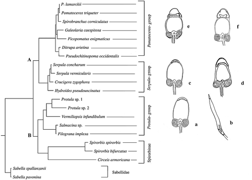
Figures & data
Figure 1. Hydroides dianthus. A, section showing segments full of male germinal cells. B, male germinal cells in various phases of maturation. C, early spermatids. er, endoplasmic reticulum; pg, proacrosomal granules. D, late spermatid. n, nucleus; a, acrosome; m, mitochondria. E, spermatozoon in the last phases of the spermioistogenesis. n, nucleus; a, acrosome; m, mitochondria; f, flagellum. F, spermatozoon. Note the indentations (arrow) in the lower part of the acrosome (a). n, nucleus; m, mitochondria; f, flagellum. G, section at the level of the proximal centriole (pc). m, mitochondria. Scale bars: A, 25 μm. B, 3 μm. C,D, 1 μm. E,F, 0.9 μm. G, 1.5 μm.
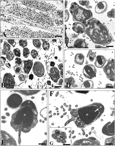
Figure 2. Serpula vermicularis. A, section showing a portion of the coelomic cavity full of male germinal cells in various phases of maturation. B, male germinal cells in various phases of maturation. sc, spermatocytes; sp, spermatozoa. C, spermatids. n, nucleus; a, acrosome; m, mitochondria. D, spermatozoon in which the condensation of the nucleus (n) is not yet complete (arrow). a, acrosome; m, mitochondria. E, spermatozoon. n, nucleus; a, acrosome; m, mitochondria, f, flagellum. F, picture at high magnification of the acrosome (a). n, nucleus. G, section at level of the flagella. Scale bars: A, 20 μm. B, 5 μm. C, 1.5 μm. D, 0.6 μm. E, 1 μm. F, 170 nm. G, 0.5 μm.
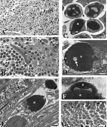
Figure 3. Vermiliopsis infundibulum. A, section showing a portion of the coelomic cavity full of male germinal cells in various phases of maturation. B, male germinal cells in various phases of maturation. sc, spermatocytes; sp, spermatozoa. C, late spermatid. n, nucleus; a, acrosome; m, mitochondria. D, spermatozoon. n, nucleus; a, acrosome; m, mitochondria; f, flagellum; pc, proximal centriole. Scale bars: A, 15 μm. B, 5 μm. C, 0.4 μm. D, 0.7 μm.
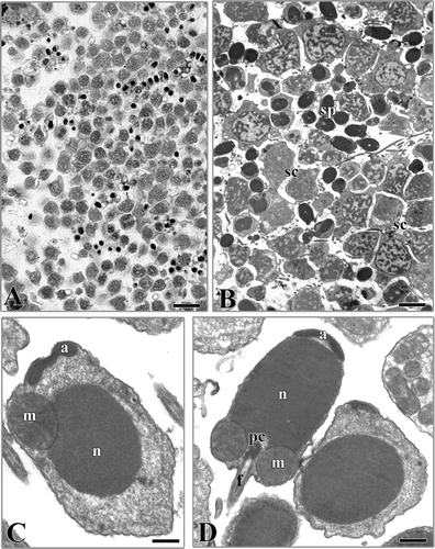
Table I. Some spermatozoa and developmental features of the up to now investigated serpulids. NF, non-feeding larva; F, feeding larva; ?, unknown
Figure 4. The spermatozoa morphology of the up to now investigated serpulid species is superimposed on the most parsimonious tree obtained, with permission, from Lehrke et al. (Citation2007). a, Vermiliopsis infundibulum (present paper); b, Salmacina sp. (redrawn, with permission, from Rouse Citation1996); c, Serpula vermicularis (present paper); d, Hydroides dianthus (present paper); e, Spirobranchus tetraceros (redrawn, with permission, from Selim et al. Citation2005); f, Galeolaria caespitosa (redrawn, with permission, from Jamieson & Rouse Citation1989).