1,926
Views
12
CrossRef citations to date
0
Altmetric
Review

Leukocyte differential counts and morphology from twelve European lizards

, , , , &
Pages 418-426 | Received 09 Aug 2010, Accepted 17 Feb 2011, Published online: 20 Apr 2011

Figures & data

Figure 1. Heterophils, eosinophils and basophils of cells of the Laceta bilineata, L. viridis, L. trilineata, L. oxycephala, Timon lepidus and Zootoca vivipara (May–Grünwald/Giemsa stain). Scale bar: 10 μm.
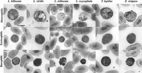
Figure 1. Heterophils, eosinophils and basophils of cells of the Laceta bilineata, L. viridis, L. trilineata, L. oxycephala, Timon lepidus and Zootoca vivipara (May–Grünwald/Giemsa stain). Scale bar: 10 μm.

Figure 2. Heterophils, eosinophils and basophils of cells of the Podarcis tiliguerta, P. muralis, P. melisellensis, P. sicula, P. bocagei, and Algyroides nigropunctatus (May–Grünwald/Giemsa stain). Scale bar: 10 μm.
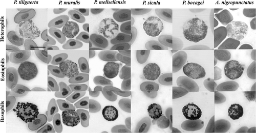
Figure 2. Heterophils, eosinophils and basophils of cells of the Podarcis tiliguerta, P. muralis, P. melisellensis, P. sicula, P. bocagei, and Algyroides nigropunctatus (May–Grünwald/Giemsa stain). Scale bar: 10 μm.

Figure 3. The fourth granulocyte type (a–h), and an erythrocyte infected by haemogregarine spp. (i) (May–Grünwald/Giemsa stain); (a) Podarcis tiliguerta, (b) P. muralis, (c) P. melisellensis , (d) P. sicula, (e) Algyroides nigropunctatus, (f) Lacerta bilineata, (g) L. trilineata, and (h) L. oxycephala. Scale bar: 10 μm.
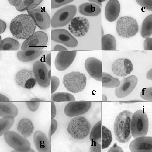
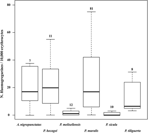
Figure 3. The fourth granulocyte type (a–h), and an erythrocyte infected by haemogregarine spp. (i) (May–Grünwald/Giemsa stain); (a) Podarcis tiliguerta, (b) P. muralis, (c) P. melisellensis , (d) P. sicula, (e) Algyroides nigropunctatus, (f) Lacerta bilineata, (g) L. trilineata, and (h) L. oxycephala. Scale bar: 10 μm.

Table I. WBC, differential leukocyte count and thrombocyte count from the blood of P. muralis

Table II. WBC, differential leukocyte count and thrombocyte count from the blood of A. nigropunctatus

Table III. WBC, differential leukocyte count and thrombocyte count from the blood of P. bocagei

Table IV. WBC, differential leukocyte count and thrombocyte count from the blood of P. melisellensis

Table V. WBC, differential leukocyte count and thrombocyte count from the blood of P. sicula

Table VI. WBC, differential leukocyte count and thrombocyte count from the blood of P. tiliguerta

Figure 4. Haemogregarine loads (N/10,000 erythrocytes) in six European lacertid lizards. Numbers above boxes indicate sample sizes.

Figure 4. Haemogregarine loads (N/10,000 erythrocytes) in six European lacertid lizards. Numbers above boxes indicate sample sizes.

Reprints and Corporate Permissions

Please note: Selecting permissions does not provide access to the full text of the article, please see our help page How do I view content?

To request a reprint or corporate permissions for this article, please click on the relevant link below:

Academic Permissions

Please note: Selecting permissions does not provide access to the full text of the article, please see our help page How do I view content?

Obtain permissions instantly via Rightslink by clicking on the button below:

If you are unable to obtain permissions via Rightslink, please complete and submit this Permissions form. For more information, please visit our Permissions help page.