Figures & data
Table I. Known associations between hydroids and anthozoans
Figure 1. Map of the sampling regions. Locations of the explored areas along the western Tyrrhenian coast: Ischia Island, shoal Secca dei Galli and S. Eufemia Gulf (black dots).
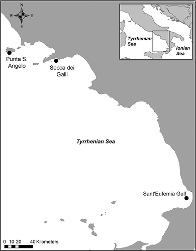
Figure 2. Description of Ectopleura sp. A, solitary polyp of Ectopleura sp. emerging from a branchlet of Antipathella subpinnata. Note the antipatharian polyps irregularly arranged on the hydroid stem. Inset: SEM close-up of the hydranth. B, solitary polyp of Ectopleura sp. emerging from a branchlet of Eunicella cavolinii. Inset: Close-up of the hydranth. C, transverse section of a hydrocaulus (hy) surrounded by the black coral (bc). Spines are visible on the golden skeleton of the coral (sk), while no divisions are found in the lumen of the hydroid. D, transverse section of a hydrocaulus (hy) surrounded by the gorgonian tissues (go). The gorgonian skeleton (sk), surrounded by the coenosarc bearing sclerites, covers the perisarc (p). Immature gonophores of Ectopleura sp. found in A. subpinnata (E) and E. cavolinii (white arrows) (F). Scale bars: B, 2 mm. A, B inset, 1 mm. A inset, 0.5 mm. C,D,F, 0.2 mm. E, 50 μm.
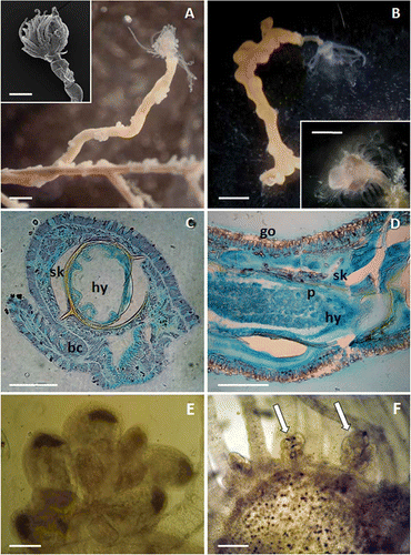
Figure 4. Relationship of Ectopleura sp. with the hosts. A, colony of Antipathella subpinnata characterised by a lax, non-arborescent ramification system. Several foreign elements (both sediment and epibionts) are visible dispersed on the branches (while arrows). B, hydroid polyp emerging perpendicularly from a black coral branchlet. C, hydrocaulus running along the black coral branchlet. D, naked hydrocaulus of Ectopleura sp. settled on Paramuricea clavata. E,F, Ectopleura sp. polyps settled on Paramuricea macrospina. G, hydroid polyp emerging perpendicularly from a branchlet of Eunicella cavolinii. H, peeled portion of E. cavolinii showing the place of settlement of the hydrocaulus. J, portion of hydrocaulus (hy) surrounded by the gorgonin layer (sk). K, perisarc of Ectopleura sp. covered by the spiny skeleton of A. subpinnata. Along the hydrocaulus diameter decreases are visible as skeleton constrictions, indicating different growing stages of the hydroid. L, SEM enlargement of the transversal section adjacent to the area of contact between Ectopleura sp. and A. subpinnata: on the outer side the black coral coenenchyme and spiny skeleton (bc), on the inner side the perisarc and the coenosarc of the hydroid (hy). M, black coral branchlet (bc) hosting Ectopleura sp. (hy). Cylindrical, pointed, irregularly arranged spines are visible near the attachment area of the hydrocaulus. Scale bars: A, 5 cm. D, 2 mm. B,C,E–H,K,M, 1 mm. J, 0.5 mm. L, 10 μm.
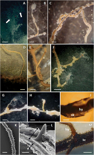
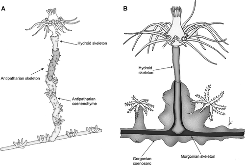
Figure 3. Scheme of the two associations. A, Ectopleura sp. on Antipathella subpinnata. B, Ectopleura sp. on Eunicella cavolinii. The distal portions of the hydrocauli have been represented as the coral tissue was removed to show the skeletons beneath.