Figures & data
Figure 1. (a) Showing a schematic diagram of the localization of the pancreas in Lissemys turtles. Sections of the splenic lobe of turtle pancreas, stained by AFT method, under light microscope showing: (a1 ) endocrine cells (A, B with arrows) with lightly stained fine cytoplasmic granules and exocrine cells (exc) with intensely stained large cytoplasmic granules. A small B-cell islet [B(I) with arrow] predominantly with B-cells, surrounded by a thin layer of greenish connective tissue (ct) (arrow) near the blood capillaries (bc); B-cells with fine blue cytoplasmic granules and A-cells with very fine orangeophilic cytoplasmic granules. B-cells in small islet [B(I)] or in small group [B(G)], and A-cells freely or in small group A(G). (b) Both B- and A-cells in small groups. One D-cell with scanty bluish-green cytoplasmic granules intermingled with orangeophilic A-cells (arrows) in the splenic lobe of the pancreas of turtle. Profuse quantity of green stained collagenous tissue (ct) associated with blood capillaries (bc). (c) One A-cell islet surrounded by green stained collagenous tissues (ct) showing mostly orangeophilic A-cells and one transforming endocrine cell (TEC (?)) with abundance of large orange–blue granules, compared to blue large granules in the exocrine cells (exc) of the pancreas. (d) One small A-islet (between the arrows) with exclusively orangeophilic granules within the epithelium of the pancreatic duct (dt) with lumen (l) surrounded by huge quantity of green stained collagenous tissue (ct). Scale bars: a1–d, 20 μm.
![Figure 1. (a) Showing a schematic diagram of the localization of the pancreas in Lissemys turtles. Sections of the splenic lobe of turtle pancreas, stained by AFT method, under light microscope showing: (a1 ) endocrine cells (A, B with arrows) with lightly stained fine cytoplasmic granules and exocrine cells (exc) with intensely stained large cytoplasmic granules. A small B-cell islet [B(I) with arrow] predominantly with B-cells, surrounded by a thin layer of greenish connective tissue (ct) (arrow) near the blood capillaries (bc); B-cells with fine blue cytoplasmic granules and A-cells with very fine orangeophilic cytoplasmic granules. B-cells in small islet [B(I)] or in small group [B(G)], and A-cells freely or in small group A(G). (b) Both B- and A-cells in small groups. One D-cell with scanty bluish-green cytoplasmic granules intermingled with orangeophilic A-cells (arrows) in the splenic lobe of the pancreas of turtle. Profuse quantity of green stained collagenous tissue (ct) associated with blood capillaries (bc). (c) One A-cell islet surrounded by green stained collagenous tissues (ct) showing mostly orangeophilic A-cells and one transforming endocrine cell (TEC (?)) with abundance of large orange–blue granules, compared to blue large granules in the exocrine cells (exc) of the pancreas. (d) One small A-islet (between the arrows) with exclusively orangeophilic granules within the epithelium of the pancreatic duct (dt) with lumen (l) surrounded by huge quantity of green stained collagenous tissue (ct). Scale bars: a1–d, 20 μm.](/cms/asset/fcd28f84-4297-4098-b223-1b0654711a6b/tizo_a_573506_o_f0001g.jpg)
Table I. Tinctorial characteristics of B- and A-cells in the endocrine pancreas of Lissemys turtles
Figure 2. (a) Insulin-immunoreactive (Insulin-IR) B-cells with brownish/deep brown reactive sites are seen in islets (I) or groups (G) in the splenic lobe of the pancreas of Lissemys turtles. (b) An enlarged view of showing deep brown reactive sites in the cytoplasm of insulin-IR B-cells (arrows) in the splenic lobe of turtle pancreas. (c) Glucagon-immunoreactive (Glucagon-IR) A-cells with deep blue reactive sites are seen in small groups (G) and in isolation in the splenic lobe of the pancreas of turtle. (d) An enlarged view of showing deep blue reactive sites in the cytoplasm of glucagon-IR A-cells (arrow) of the splenic lobe of turtle pancreas. Scale bars: a–d, 20 μm.
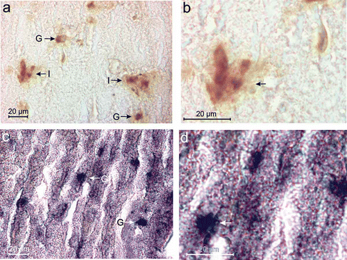
Figure 3. (a) Electron micrograph showing indented (I) or lobulated (L) nucleus with smaller secretory granules (Ssg) in the endocrine cells (endo) (arrow) compared to those of larger secretory granules (Lsg) of the exocrine cells (exc) in the splenic lobe of the pancreas of Lissemys turtles. Scale bar: 10 μm. (b) Electron micrograph showing distribution of endocrine cells (endo) mostly near the blood capillaries (bc). Secretory granules (sg) are smaller in the endocrine cells (endo) than in exocrine cells (exc). Scale bar: 10 μm.
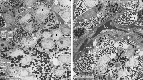
Table II. Cellular and intracellular characteristics of endocrine cells in the pancreas under TEM in Lissemys turtles
Table III. Ultrastructural characteristics of the secretory granules of the endocrine cells of the pancreas of Lissemys turtles
Figure 4. Electron micrographs of the B-cell of the endocrine pancreas of the splenic lobe of Lissemys turtles showing (a) an intact large, oval B-cell close to blood capillaries (bc), with a very large indented euchromatic nucleus (n), mitochondrial region (mt) and moderate abundance of homogenously distributed secretory granules (sg) barring the mitochondrial region. (b) An enlarged view of showing conspicuous mitochondrial (mt) region with few rough endoplasmic reticulum (RER) at the periphery. (c) Short RER is seen very close to the oval mitochondria (mt) with conspicuous cristae. (d) Golgi bodies (G) with concentric vesicles are seen surrounding a secretory granule (sg). (e) Showing secretory granules (sg) with crystalline-like electron-dense core and very wide electron-lucent peripheral halo. (f) Other secretory granules showing negligible electron-dense part and very wide halo. (g) Showing small vacuoles (V) close to the secretory granules (sg). (h) Some B-cells (?) are intermingled in the epithelium (ep) of the pancreatic duct with few endocrine cells (endo) close to the duct. Scale bars: a, 2 μm. b, 1 μm. c–f, 0.25 μm. g, 0.5 μm. h, 5 μm.
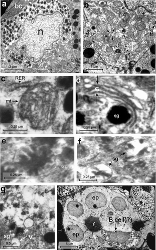
Figure 5. Electron micrographs of the A-cell of the endocrine pancreas of the splenic lobe of Lissemys turtles showing (a) an intact A-cell with elongated and indented euchromatic nucleus (n), RER region, mitochondrial region (mt), lysosomes (ly) and moderate abundance of homogeneously distributed electron-dense secretory granules (sg) barring the mitochondrial region. (b) Showing long filamentous RER, encircling the secretory granules (sg). (c) Long filamentous and branched mitochondria (mt) with inconspicuous cristae are seen in the cytoplasm. (d) Golgi apparatus (G) with concentric vesicles surrounding a secretory granule (sg) is seen. (e) Showing a multivescicular body (arrows) with numerous empty vesicles of various sizes. Scale bars: a, 2 μm. b, d, 0.25 μm. c, 1 μm. e, 0.5 μm.
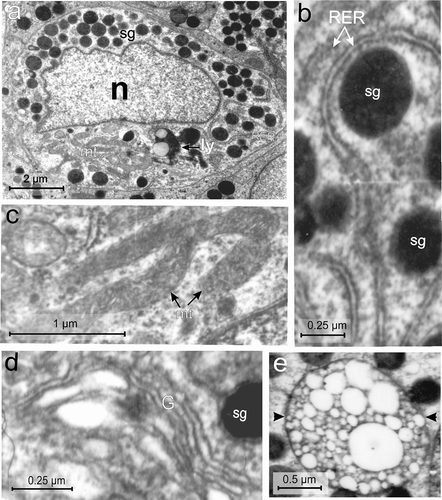
Figure 6. Electron micrographs of the D-cell of the endocrine pancreas of the splenic lobe of Lissemys turtles showing (a) an intact oval D-cell adjacent to the blood capillaries (bc), containing a large oval euchromatic nucleus (n) and moderate number of electron-dense secretory granules (sg) of various size, aggregated in small groups with a RER region. (b) Branched RER with dilated cisternae is seen. (c) Showing mitochondria (mt) with conspicuous cristae. (d) Secretory granules (sg) with wide electron-dense core and narrow electron-lucent peripheral halo are seen. Scale bars: a, 2 μm. b–d, 0.25 μm.
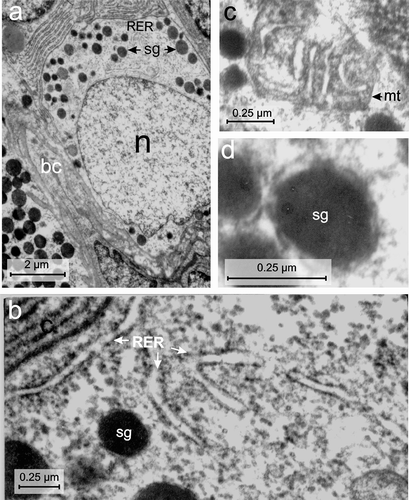
Figure 7. Electron micrographs of the PP-cell of the endocrine pancreas of the splenic lobe of Lissemys turtles showing (a) two fusiform cells (arrows) with tapering ends adjacent to the blood capillaries (bc), large euchromatic oval nucleus (n) (partly seen) and moderate abundance of small electron-dense secretory granules (sg). (b) A PP-cell is seen adjacent to an exocrine cell (exc) (arrowhead) with an intact indented nucleus (n), RER region and mitochondrial regions (mt). (c) An enlarged view of RER with dilated cisternae in the cell cytoplasm is seen. (d) A magnified view of an oval mitochondrium (mt) showing conspicuous cristae adjacent to few secretory granules (sg). (e) The secretory granules (sg) with a wide electron-dense core and a thin layer of peripheral halo are seen. Scale bars: a, b, 2 μm. c, 1 μm. d, 0.5 μm. e, 0.25 μm.
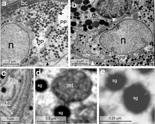
Figure 8. Electron micrographs of the Vth-cell type of the endocrine pancreas of the splenic lobe of the Lissemys turtles showing (a) an intact triangular in shape, located adjacent to exocrine cells (exc) with indented hyperchromatinic nucleus (n), containing a prominent nucleolus (nu) near the nuclear membrane, mitochondrial region (mt) and abundance of secretory granules (sg) aggregated in two corners of the cell. (b) Oval and elongated mitochondria (mt) with conspicuous cristae and vacuoles (v) are seen in the cell cytoplasm. (c) Pear-shaped secretory granules (sg) showing wide electron-dense core with a narrow peripheral halo. Scale bars: a, 2 μm. b, 0.5 μm. c, 0.125 μm.
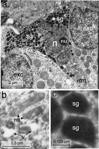
Figure 9. Electron micrographs of the VIth-cell type of the endocrine pancreas of the splenic lobe of Lissemys turtles showing (a) a very small, pyramidal in shape, with a large euchromatic indented nucleus (n), mitochondrial region (mt) and scanty secretory granules (sg), aggregated in small groups. (b) Several round/oval mitochondria with conspicuous cristae are seen. (c) Showing oval secretory granules (sg) with wide electron-dense core and narrow peripheral halo. Scale bars: a, 2 μm. b, 1 μm. c, 0.25 μm.
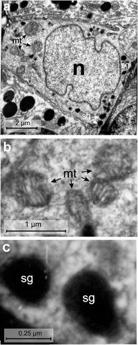
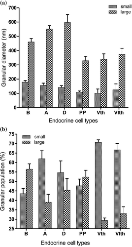
Figure 10. (a) Histograms (representing mean ± SE values) showing diameters (nm) of small and large secretory granules of different endocrine cells (B, A, D, PP, Vth and VIth) in the splenic lobe of the pancreas of Lissemys turtles. (b) Histograms (representing mean ± SE values) showing populations (%) of small and large secretory granules of different endocrine cell types (B, A, D, PP, Vth and VIth) in the splenic lobe of the pancreas of Lissemys turtles.
