519
Views
8
CrossRef citations to date
0
Altmetric
Articles

First record of Kinnecaris (Copepoda: Harpacticoida: Parastenocarididae) from Turkey and Thailand; description of three new species and emended definition of the genus

&
Pages 69-94 | Received 10 Sep 2014, Accepted 15 Dec 2014, Published online: 06 Feb 2015

Figures & data

Figure 1. Kinnecaris xanthi sp. nov. A, male, habitus, lateral view. B, female, habitus, lateral view (sensillar pattern omitted). Scale bar: 50 µm.
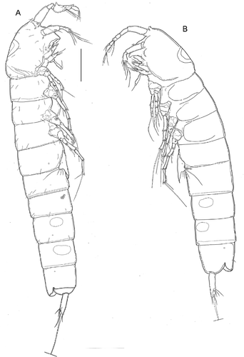
Figure 1. Kinnecaris xanthi sp. nov. A, male, habitus, lateral view. B, female, habitus, lateral view (sensillar pattern omitted). Scale bar: 50 µm.

Figure 2. Kinnecaris xanthi sp. nov. A, male, fourth and fifth urosomites, anal somite, anal operculum and caudal ramus, lateral view. B, male, anal somite, anal operculum and caudal rami, dorsal view. C, male, spermatophore. D, male, rostrum and antennule, dorsal view. E, male, antennule, outer view, schematic, main armature omitted. F, male, antennule, ventral view (armature partly omitted, insertion point of setae marked by circles). G, male, antenna. H, male, labrum. I, male, mandible. J, male, maxillule. K, male, maxilla. L, male, maxilliped. Scale bar: 50 µm.
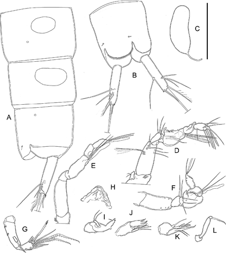
Figure 2. Kinnecaris xanthi sp. nov. A, male, fourth and fifth urosomites, anal somite, anal operculum and caudal ramus, lateral view. B, male, anal somite, anal operculum and caudal rami, dorsal view. C, male, spermatophore. D, male, rostrum and antennule, dorsal view. E, male, antennule, outer view, schematic, main armature omitted. F, male, antennule, ventral view (armature partly omitted, insertion point of setae marked by circles). G, male, antenna. H, male, labrum. I, male, mandible. J, male, maxillule. K, male, maxilla. L, male, maxilliped. Scale bar: 50 µm.

Figure 3. Kinnecaris xanthi sp. nov. A, male, leg 1. B, male, leg 2. C, male, leg 3. D, male, leg 3 (variability). E, male, leg 4, dorsal view. F, male, leg 4, ventral view. G, male, leg 4 (variability). H, male, first and second urosomites, leg 5, leg 6, ventral view. Scale bar: 50 µm.
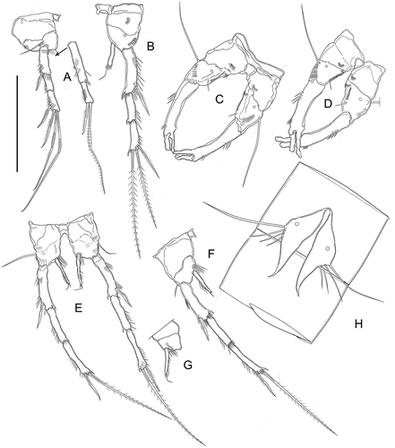
Figure 3. Kinnecaris xanthi sp. nov. A, male, leg 1. B, male, leg 2. C, male, leg 3. D, male, leg 3 (variability). E, male, leg 4, dorsal view. F, male, leg 4, ventral view. G, male, leg 4 (variability). H, male, first and second urosomites, leg 5, leg 6, ventral view. Scale bar: 50 µm.

Figure 4. Kinnecaris xanthi sp. nov. A, female, fourth and fifth urosomites, anal somite, anal operculum and caudal ramus, lateral view. B, female, first urosomite, leg 5, genital double-somite and genital field, ventral view. C, female, antennule, dorsal view. D, female, leg 2. E, female, leg 3. F, female, leg 4. Scale bar: 50 µm.
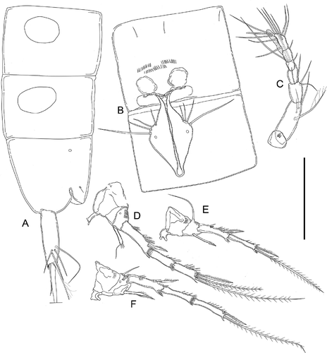
Figure 4. Kinnecaris xanthi sp. nov. A, female, fourth and fifth urosomites, anal somite, anal operculum and caudal ramus, lateral view. B, female, first urosomite, leg 5, genital double-somite and genital field, ventral view. C, female, antennule, dorsal view. D, female, leg 2. E, female, leg 3. F, female, leg 4. Scale bar: 50 µm.

Figure 5. Kinnecaris draconis sp. nov. A, male, habitus, lateral view. B, female, habitus, lateral view. Scale bar: 50 µm.
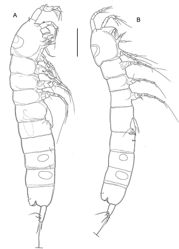
Figure 5. Kinnecaris draconis sp. nov. A, male, habitus, lateral view. B, female, habitus, lateral view. Scale bar: 50 µm.

Figure 6. Kinnecaris draconis sp. nov. A, male, fourth and fifth urosomites, anal somite, anal operculum and caudal rami, dorsal view. B, male, spermatophore. C, male, antennule, dorsal view. D, male, antenna. E, male, mandible. F, male, maxillule. G, male, maxilla. H, male, maxilliped. I, male, leg 1. J, male, leg 2. K, male, leg 3, lateral view (variability). L, male, leg 3. M, male, leg 4. Scale bar: 50 µm.
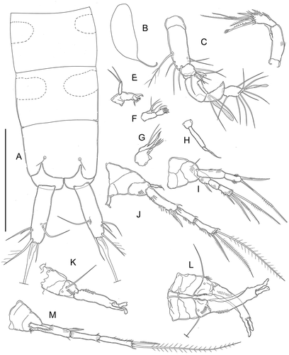
Figure 6. Kinnecaris draconis sp. nov. A, male, fourth and fifth urosomites, anal somite, anal operculum and caudal rami, dorsal view. B, male, spermatophore. C, male, antennule, dorsal view. D, male, antenna. E, male, mandible. F, male, maxillule. G, male, maxilla. H, male, maxilliped. I, male, leg 1. J, male, leg 2. K, male, leg 3, lateral view (variability). L, male, leg 3. M, male, leg 4. Scale bar: 50 µm.

Figure 7. Kinnecaris draconis sp. nov. A, male, leg 5. B, female, antennule, ventral view. C, female, leg 3. D, female, leg 4. E, female, first urosomite, leg 5, genital double-somite and genital field, ventral view. F, female, fourth and fifth urosomites, anal somite, anal operculum and caudal ramus, lateral view. G, female, fourth and fifth urosomites, anal somite, anal operculum and caudal ramus, dorsal view. Scale bar: 50 µm.
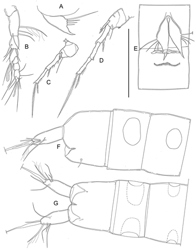
Figure 7. Kinnecaris draconis sp. nov. A, male, leg 5. B, female, antennule, ventral view. C, female, leg 3. D, female, leg 4. E, female, first urosomite, leg 5, genital double-somite and genital field, ventral view. F, female, fourth and fifth urosomites, anal somite, anal operculum and caudal ramus, lateral view. G, female, fourth and fifth urosomites, anal somite, anal operculum and caudal ramus, dorsal view. Scale bar: 50 µm.

Figure 8. Kinnecaris iulianae sp. nov. A, male, habitus, lateral view. B, female, habitus, lateral view (sensillar pattern omitted). Scale bar: 50 µm.
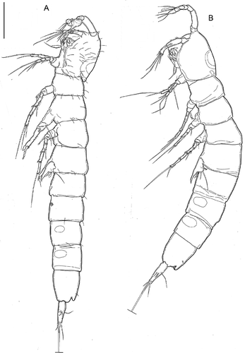
Figure 8. Kinnecaris iulianae sp. nov. A, male, habitus, lateral view. B, female, habitus, lateral view (sensillar pattern omitted). Scale bar: 50 µm.

Figure 9. Kinnecaris iulianae sp. nov. A, male, double dorsal cuticular window on cephalothorax, lateral view. B, male, leg 5, leg 6, first to fourth urosomites, ventral view. C, male, fourth and fifth urosomites, anal somite, anal operculum and caudal rami, ventral view (square insert with detail of pitted cuticle). D, male, fourth and fifth urosomites, anal somite, anal operculum and caudal ramus, lateral view. E, male, spermatophore. F, male, antennule, dorsal view. G, male, antenna. H, male, mandible. I, male, maxillule. J, male, maxilla. K, male, maxilliped. Scale bar: 50 µm.
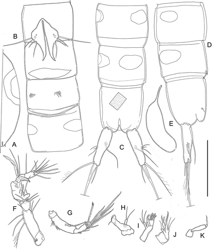
Figure 9. Kinnecaris iulianae sp. nov. A, male, double dorsal cuticular window on cephalothorax, lateral view. B, male, leg 5, leg 6, first to fourth urosomites, ventral view. C, male, fourth and fifth urosomites, anal somite, anal operculum and caudal rami, ventral view (square insert with detail of pitted cuticle). D, male, fourth and fifth urosomites, anal somite, anal operculum and caudal ramus, lateral view. E, male, spermatophore. F, male, antennule, dorsal view. G, male, antenna. H, male, mandible. I, male, maxillule. J, male, maxilla. K, male, maxilliped. Scale bar: 50 µm.

Figure 10. Kinnecaris iulianae sp. nov. A, male, leg 1. B, male, leg 2. C, male, leg 3, D, male, leg 4, lateral view. E, male, leg 4. F, male, leg 5. G, male, first urosomite and P5 (variability), ventral view. H, male, leg 5 (variability). Scale bar: 50 µm.
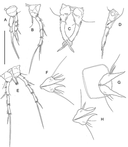
Figure 10. Kinnecaris iulianae sp. nov. A, male, leg 1. B, male, leg 2. C, male, leg 3, D, male, leg 4, lateral view. E, male, leg 4. F, male, leg 5. G, male, first urosomite and P5 (variability), ventral view. H, male, leg 5 (variability). Scale bar: 50 µm.

Figure 11. Kinnecaris iulianae sp. nov. A, female, fifth urosomites, anal somite, anal operculum and caudal ramus, lateral view. B, female, fourth and fifth urosomites, anal somite, anal operculum and caudal rami, dorsal view. C, female, genital double-somite, genital field, P5, ventral view. D, female, antennule. E, female, leg 2. F, female, leg 3. G, female, leg 4. H, female, leg 5. I, female, leg 5 (variability). J, female, genital field, ventral view. Scale bar: 50 µm.
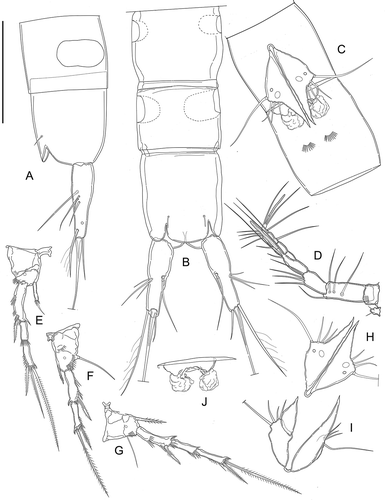
Figure 11. Kinnecaris iulianae sp. nov. A, female, fifth urosomites, anal somite, anal operculum and caudal ramus, lateral view. B, female, fourth and fifth urosomites, anal somite, anal operculum and caudal rami, dorsal view. C, female, genital double-somite, genital field, P5, ventral view. D, female, antennule. E, female, leg 2. F, female, leg 3. G, female, leg 4. H, female, leg 5. I, female, leg 5 (variability). J, female, genital field, ventral view. Scale bar: 50 µm.

Figure 12. Kinnecaris iulianae sp. nov. Pictures taken with phase-contrast microscope at 40x. A, male, fourth and fifth urosomites, anal somite, anal operculum and caudal rami, ventral view. B, male, antennule, ventral view. C, female, legs 1 to 5, lateral view. D, female, fifth urosomites, anal somite, anal operculum and caudal rami, ventral view. Scale bar: 50 µm.
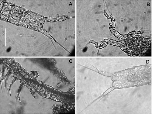
Figure 12. Kinnecaris iulianae sp. nov. Pictures taken with phase-contrast microscope at 40x. A, male, fourth and fifth urosomites, anal somite, anal operculum and caudal rami, ventral view. B, male, antennule, ventral view. C, female, legs 1 to 5, lateral view. D, female, fifth urosomites, anal somite, anal operculum and caudal rami, ventral view. Scale bar: 50 µm.

Figure 13. Kinnecaris lyncaea (Cottarelli and Bruno, Citation1994). A, male, antennule, dorsal view. B, male, first and second urosomites, leg 5 and leg 6, ventral view. Scale bar: 50 µm.
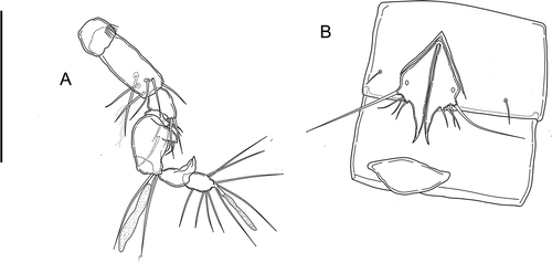
Figure 13. Kinnecaris lyncaea (Cottarelli and Bruno, Citation1994). A, male, antennule, dorsal view. B, male, first and second urosomites, leg 5 and leg 6, ventral view. Scale bar: 50 µm.

Reprints and Corporate Permissions

Please note: Selecting permissions does not provide access to the full text of the article, please see our help page How do I view content?

To request a reprint or corporate permissions for this article, please click on the relevant link below:

Academic Permissions

Please note: Selecting permissions does not provide access to the full text of the article, please see our help page How do I view content?

Obtain permissions instantly via Rightslink by clicking on the button below:

If you are unable to obtain permissions via Rightslink, please complete and submit this Permissions form. For more information, please visit our Permissions help page.