Figures & data
Figure 1. Map of the dive sites where Chalinula nematifera was observed.
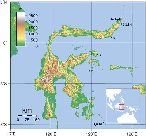
Table I. Areas and dive sites where Chalinula nematifera was recorded associated with hard corals.
Figure 2. Chalinula nematifera from Bangka Island: A–C, Acropora necrosis (white band) in the area in contact with the sponge; arrow in (A) points the starting point where the plastic wire was tied to measure sponge growth; D, white area of dead Acropora on a branch in contact with a covered colony; E, sponge invading the encrusting coral Pachyseris sp. and Seriatopora sp.; F, filaments of mucus (arrow) produced by the sponge.
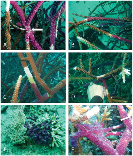
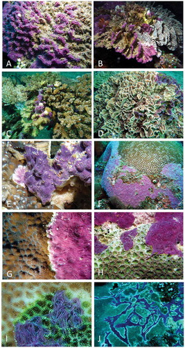
Figure 3. Chalinula nematifera overgrowing hard corals: A, unidentified species; B, Pectinia sp.; C, Pocillopora sp.; D, Pectinia sp.; E, Mycedium elephantothus; F, Platygyra daedalea; G, Echinopora sp.; H, Favia sp.; I, Goniastrea sp.; J, Faviidae. Note particularly in (H–J) the white areas of necrosis, and the sponge spreading out over living portions of coral in (C–E).