Figures & data
Table I. Localities and coordinates where the species whose stridulatory organs have been described in this study were sampled.
Figure 1. Scanning electron microscopy photographs of the head of Aphaenogaster senilis and the stridulatory organ of Myrmica sabuleti, showing the way measurements were taken for the morphometric study: cephalic width and pars stridens length (vertical) and width (horizontal).
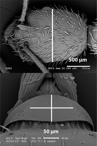
Table II. Average values of the morphometric variables of the studied myrmicine species and their standard deviation (SD).
Figure 2. Scanning electron microscopy photograph of the stridulatory organs of Aphaenogaster striativentris.
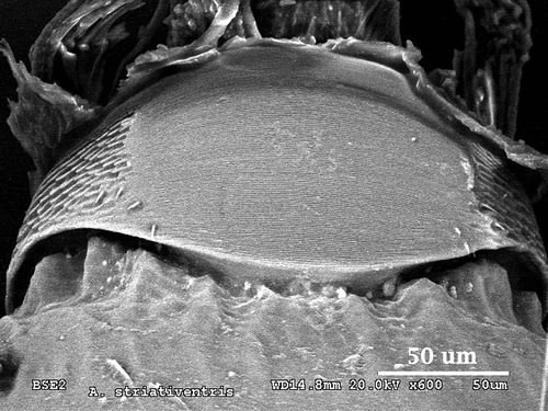
Figure 3. Scanning electron microscopy photographs of the stridulatory organs of Goniomma hispanicum (above) and detail of the toothed pillars (below).
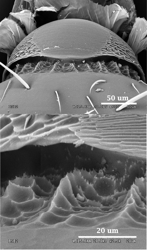
Figure 4. Scanning electron microscopy photograph of the stridulatory organs of Goniomma blanci.
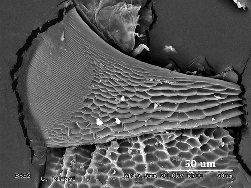
Figure 5. Scanning electron microscopy photograph of the stridulatory organs of Oxyopomyrmex saulcyi.
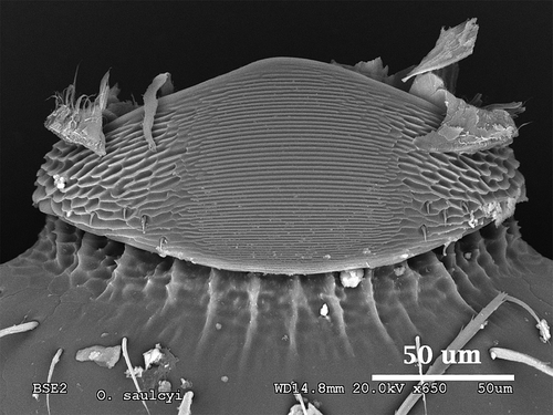
Figure 6. Scanning electron microscopy photographs of the stridulatory organs of Pheidole pallidula (above) and detail of the plectrum (below).
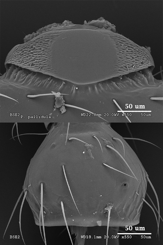
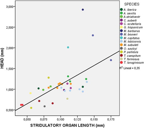
Figure 7. Scatter plot showing the relationship between the length of the pars stridens of the stridulatory organs and the cephalic width of the individual in speciemens of the 16 species considered for the analysis. Data for the listed species are shown in different colours.