Figures & data
Figure 1. Location of the study area: A, Southern part of the Venice lagoon (Val di Brenta); B, Marinetta lagoon; C, Barbamarco lagoon. Dots represent the location of the sampling stations. Dotted lines are the Po riverbed branches. Shaded areas represent the location of ports, harbours and marinas.
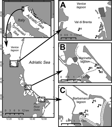
Table I. Characterisation of sampling sites where specimens of Polydora cornuta were collected in the three North Adriatic lagoons. The number of individuals is the sum of three replicates. The number of mature females includes the females hatching egg capsules.
Figure 2. Polydora cornuta. A, anterior end, dorsal view; a = occipital antenna; B, chaetiger 5 from right side, dorsal view; sp = modified spines; cc = companion chaetae strictly adhering to convex side of the spine; C, chaetae from chaetiger 5: from left to right, unworn posterior spine; unworn posterior companion chaeta; worn anterior companion chaeta on convex (left) and concave (right) side; D, notopodial chaetae from chaetiger 20: s = short capillaries, w = winged capillaries, lc = long capillaries, the square shows the detail of the winged terminal part of the chaeta; E, hooded hook from posterior chaetigers; F, posterior end and pygidium. Scale bars: A = 1.5 mm; B, D = 100 mm; C = 50 mm; E = 30 mm; F = 1 mm.
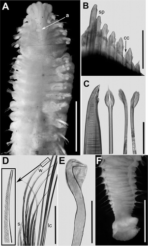
Figure 3. Polydora cornuta. A, relationship between size of individuals (width of chaetiger 5) and number of spines in the same chaetiger (linear model: y = 3.24 + x·4.01; r = 0.853; P < 0.0001; N = 45); B, relationship between size of individuals (width of chaetiger 5) and maximum number of hooded hooks in posterior segments (linear model: y = 2.46 + x·6.88; r = 0.875; P < 0.0001; N = 41).
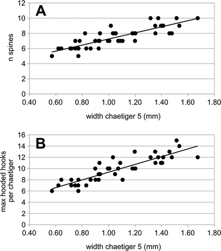
Figure 4. Polydora cornuta. A, mud tube with egg capsules attached to inner wall; B, early stage of a three-chaetiger larva extracted from a capsule: yg = yolky globules. Scale bars: A = 1 mm; B = 150 mm.
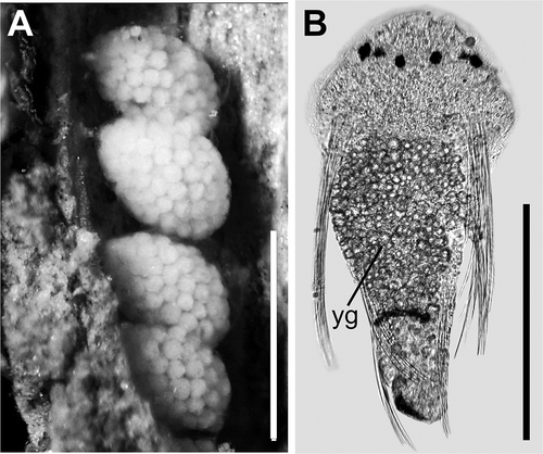
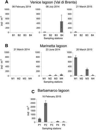
Figure 5. Polydora cornuta mean densities measured at each sampling station at each sampling time. A, Venice lagoon (Val di Brenta); B, Marinetta lagoon; C, Barbamarco lagoon. Error bars are standard errors (n = 3).