Figures & data
Figure 1. Hypophthalmichthys molitrix, 2 days after fertilization. Light microscopy micrograph of a transverse section across the developing inner ear, showing the first appearance of the otic vesicle (av), containing the otolith primordia (op) and the stato-acoustic ganglion (sg). br, brain. 550×.
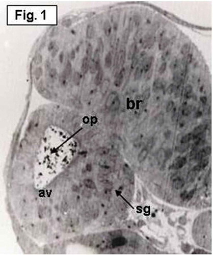
Figure 2. Hypophthalmichthys molitrix, 3 days after fertilization. Light microscopy micrograph of a transverse section across the developing inner ear, showing first appearance of the saccular otolith (so) on an accumulation of polygonal cells, representing the saccular macula (sm). Note the stato-acoustic ganglion (sg) behind the saccular macula. br, brain. 350×.
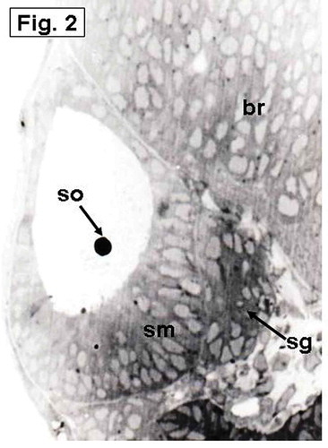
Figure 3. Hypophthalmichthys molitrix, 4 days after fertilization Light microscopy micrograph of a transverse section across the developing inner ear, showing arrangement of the saccular macular cells (sm) in an oblique position overlain by the saccular otolith (so). br, brain. 370×.
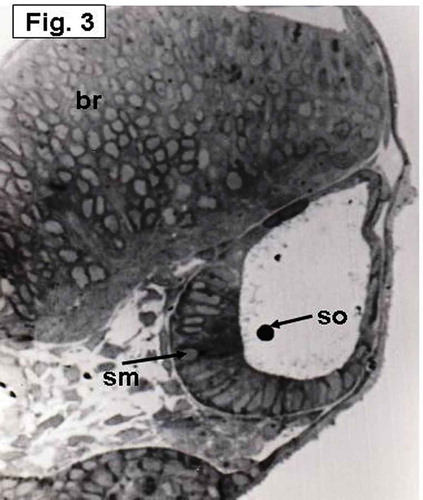
Figure 4. Hypophthalmichthys molitrix, 4 days after fertilization. Light microscopy micrograph of a transverse section across the developing inner ear, showing establishment of the utricular macula (um) which overlain by a small utricular otoloith (uo). br, brain. 355×.
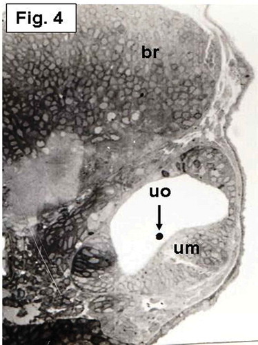
Figure 5. Hypophthalmichthys molitrix, 3 days after hatching. Light microscopy micrograph of a transverse section across the developing inner ear, showing the beginning of differentiation of the saccular macular epithelium (sm) that positioned in a vertical position and restricted to the medial wall. The saccular macula (sm) overlain by an elongated saccular otolith (so) which covers the middle part of the macula. br, brain; asc, anterior semicircular canal; hsc, horizontal semicircular canal. 200×.
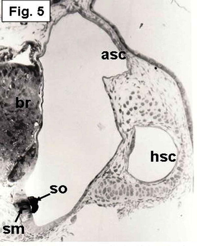
Figure 6. Hypophthalmichthys molitrix, 7 days after hatching. Light microscopy micrograph of a transverse section through the developing inner ear showing the saccular macula (sm) which is more invaginated toward the ventro-medial wall. The saccular otolith (so) increased in size and covered most of the saccular epithelial cells. br, brain. 250×.
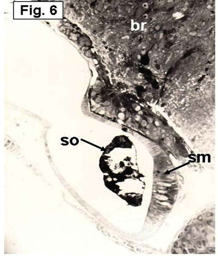
Figure 7. Hypophthalmichthys molitrix, 7 days after hatching. Light microscopy micrograph of a transverse section through the developing inner ear showing differentiation of the saccular macula into transitional epithelium (te) and sensory epithelium that differentiate into hair cells (arrows) and supporting cells (sc). so, saccular otolith. 300×.
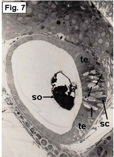
Figure 8. Hypophthalmichthys molitrix, 7 days after hatching. TEM micrograph of the saccular sensory epithelium, showing supporting cells (sc) and a hair cell (hc) which possesses a hair bundle (hb) that planted in a cuticular plate (c). Secretory materials, like cytoplasmic extrusion (ce) and empty vesicles (arrows), seemed to be librated from supporting cells (sc). Microridges (mr) can be seen on the apical surface of a supporting cell. j, junctional complex; st, stereocilia of the hair bundle. 8300×.
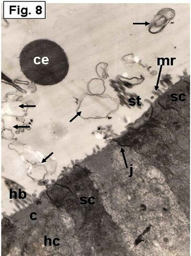
Figure 9. Hypophthalmichthys molitrix, 7 days after hatching. TEM micrograph of the saccular sensory epithelium, showing the otolith (ot) covering hair cells (hc) and supporting cells (sc). Secretory materials like numerous vesicles (arrows), cytoplasmic extrusion (ce) and electron dense granules (eg) can be seen between the otolith and the sensory epithelium. hb, hair bundle. 6300×.
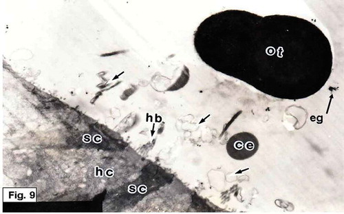
Figure 10. Hypophthalmichthys molitrix, 7 days after hatching. TEM micrograph of the saccular sensory epithelium, showing cytoplasmic extrusion (ce) projecting from supporting cell (sc) and still in contact with its apical surface. Secretory materials, as vesicular bodies (vb) containing small spherules, seemed to be librated from supporting cell. Note the microridges (mr) of the supporting cell. 17,000×.
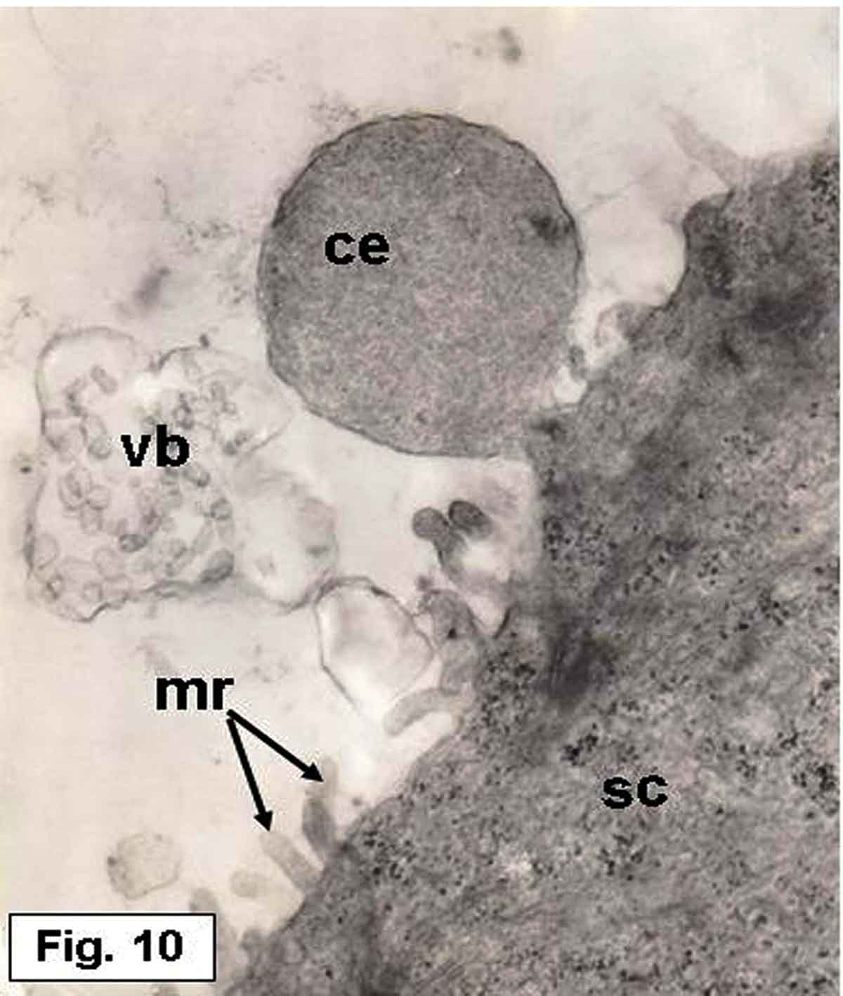
Figure 11. Hypophthalmichthys molitrix, 7 days after hatching. TEM micrograph of the saccular sensory epithelium, showing an accumulation of various kinds of secretory materials over the saccular sensory epithelium; empty vesicles (ev), electron dense granules (arrows). Note the stereocilia (st) of a hair bundle projecting from the cuticular plate (asterisk) of the hair cell (hc). The supporting cells (sc) provided with microridges (mr) on their apical surfaces. 8500×.
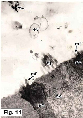
Figure 12. Hypophthalmichthys molitrix, 7 days after hatching. TEM micrograph of a sensory hair cell (hc), showing the stereocilia (st) which is planted in a cuticular plate (arrow). Secretory materials, as vesicular bodies (vb) containing numerous spherules and other vesicles (asteriske), are also seen. 15,000×.
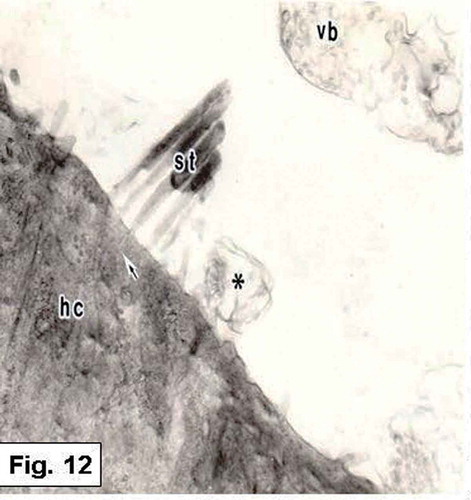
Figure 13. Hypophthalmichthys molitrix, 7 days after hatching. TEM micrograph of a sensory hair cell, showing numerous mitochondria (arrows) in the supranuclear part of the hair cell (hc). c, cuticular plate; hb, hair bundle. 16,000×.
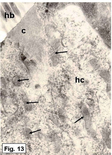
Figure 14. Hypophthalmichthys molitrix, adult specimen. SEM micrograph of the saccular sensory epithelium, showing the first type of the sensory hair bundles, each one consisted of numerous short stereocilia (st) and a kinocilium (k) which is twice as long as the longest stereocilium. 3900×.
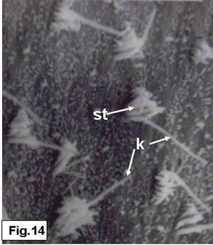
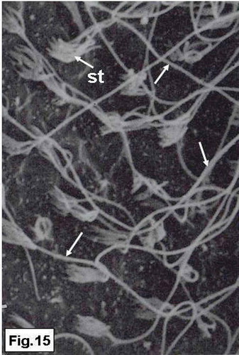
Figure 15. Hypophthalmichthys molitrix, adult specimen. SEM micrograph of the saccular sensory epithelium, showing the second type of the sensory hair bundles, each one consisted of a small number of short stereocilia (st) and very long kinocilium (arrows). 3850×.