
Figures & data
Figure 1. Field photos of the sampling sites. A: Sampling site on the South African (eastern) side of the Drakensberg escarpment, showing the cascading roots of the plants from which the sample was taken; B: Sampling site on the Lesotho (western) side of the Drakensberg escarpment, indicating algal growth in the stream from which scrapes were made.
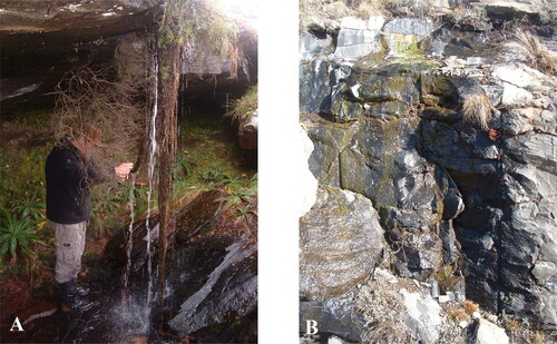
Figure 2. Light microscope images of Actinodontum lomaense cells from the Drakensberg escarpment. Scale bars = 20 µm.
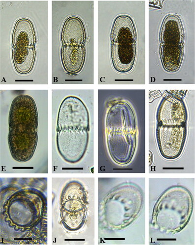
Figure 3. Scanning electron microscope images of Actinodontum lomaense cells from the Drakensberg escarpment. Scale bars: 20 µm.
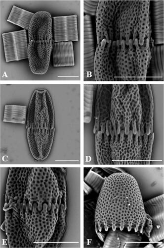
Table 1. Cell dimensions (in µm) found in the samples from the Drakensberg.