Figures & data
Figure 1. Plants of Iris ensata. (A) Plants in the field; (B–C) Symptoms; (D) Pathogenicity on detached leaves.
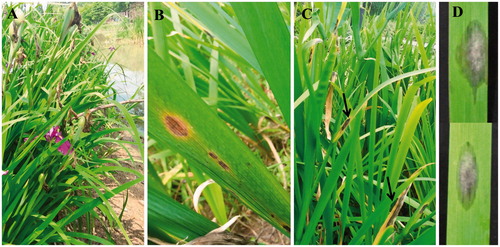
Figure 2. Morphological features of the fungus described in this work. (A) Colony characterization on PDA after 7 d. (B–D) Sporulation patterns on PCA. (E) Conidia (scale bars: B–E = 20 µm).
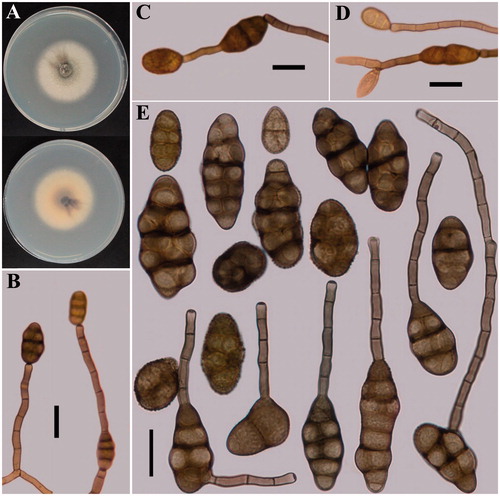
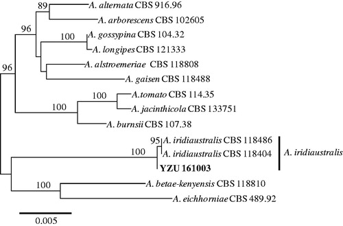
Figure 3. Maximum likelihood tree of A. iridiaustralis, generated from a combined analysis of ITS, gpd, endoPG, and RPB2 gene datasets. Numbers above the branches indicate bootstrap values (≥60%) obtained for 1000 replicates. The scale bar indicates the number of substitutions per position.