Figures & data
Figure 1. Inhibition of P. palmivora by P. aeruginosa RS1 on a V8 agar plate (A) and control growth of the mold without the bacteria (B). Microscopic observation (400×) of the hyphae of P. palmivora growing with (C) and without (D) P. aeruginosa RS1.
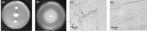
Figure 2. Inhibition percentage against P. palmivora and absorbance at OD660 nm of P. aeruginosa RS1 in LB medium (pH 7) at 37 °C, at time points measured every 3 h, 24 h.
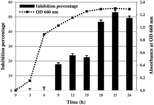
Table 1. Percentage of inhibition by culture filtrate from P. aeruginosa RS1 at different variable factors.
Figure 3. Chromatogram from DEAE-cellulose column chromatography indicates the absorbance at OD280 nm of the proteins eluting from the column using gradient concentrations of NaCl.
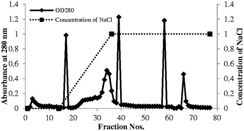
Figure 4. Inhibition of P. palmivora by eluted proteins from DEAE-cellulose column chromatography. P. palmivora grew on a V8 agar plate: control without protein (A), with protein fraction 9–10 (B), and with protein fraction 33–34.
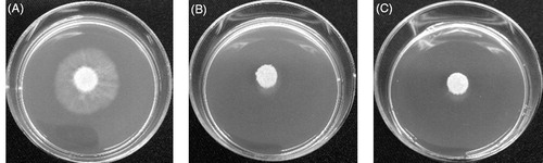
Figure 5. SDS-PAGE of partial purification of the antifungal proteins from the ammonium sulfate precipitation and anion exchange chromatography steps. The labels 9–10 and 33–34 refer to the proteins from DEAE fractions 9–10 and 33–34, respectively.
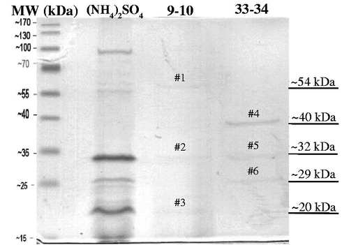
Table 2. Summary for purification of the antifungal protein from P. aeruginosa RS1.
Table 3. Identification of each protein band from anion exchange chromatography fractions 9–10 and 33–34.
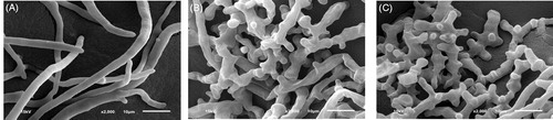
Figure 6. Morphology of P. palmivora under scanning electron microscope (2000×) after incubation for 2 days without (A) and with the purified antifungal protein from DEAE-cellulose column chromatography at fractions 9–10 (B) and 33–34 (C).