
Figures & data
Figure 1. Symptoms of gray mold disease on C. japonicum. (A, B) Gray mold symptom of C. japonicum at a natural habitat; (C) Colony morphology of B. cinerea on PDA media; (D) Conidia of B. cinerea, isolated from C. japonicum. Red bar presents 50 μm.
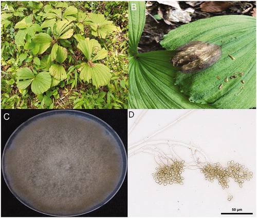
Table 1. Comparison of morphological characteristics of gray mold fungus isolated from Cypripedium japonicum.
Figure 2. Phylogenetic tree obtained through the maximum-likelihood and maximum parsimony analyses using MEGA 10 program base on the (A) ITS, (B) HSP60, and (C) G3PDH sequence of the isolates of B. cinerea and that of other B. cinerea isolates were retrieved from GenBank.
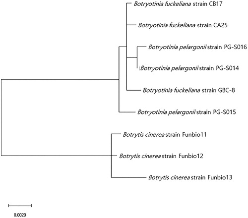
Table 2. List and information of primers for fungal identification.