Figures & data
Table 1. Primers used for amplification.
Table 2. PCR conditions used for amplification.
Table 3. Sequences used in this study.
Figure 1. Phylogenetic tree of combined LSU, tef1-α, rpb2, rpb1, and SSU sequence data inferred from 17 taxa and 5257 sites under the GTR (general time reversible) + GAMMA model of nucleotide substitution. Numerical values at the nodes indicate maximum likelihood bootstrap support (blue), maximum parsimony bootstrap support (yellow), and posterior probabilities (black). Only values greater than 70% (maximum likelihood and maximum parsimony) and 0.95 (Bayesian analysis) are indicated. Name in red indicates new species. Names in blue indicate new collections. Tree is artificially rooted to Monascella botryosa (CBS 233.85) and Warcupia terrestris (CBS 891.69) taxa.
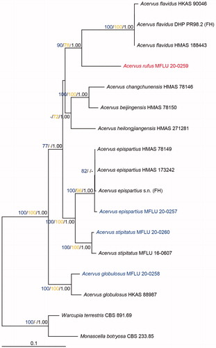
Figure 2. Acervus epispartius (HKAS 107301). (a–d) Typical mature specimens. (e) Receptacle surface of pileus in 5% KOH. (f) Asci and paraphyses in 5% KOH. (g, h) Paraphyses in Congo red. (i, j) Asci. (j Asci in Congo red.). (k, l) Apexes of asci. (k Apex in Congo red. l Apex in Cotton blue.). m-p Ascospores. Scale bars: e–f = 50 μm. g–h = 20 μm. i–j = 30 μm. k–p = 5 μm.
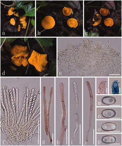
Figure 3. Acervus globulosus (MFLU 20-0258). (a–f) Typical mature specimens. (g, h) Receptacle surface of pileus. (i) Asci and paraphyses. (j–l) Asci (l Asci in Melzer’s reagent.). (m–p) Apexes of asci (m–o Apexes in Congo red.). (q–u) Ascospores. Scale bars: c = 1500 μm. d–f = 1000 μm. g–l = 30 μm. m–u = 5 μm.
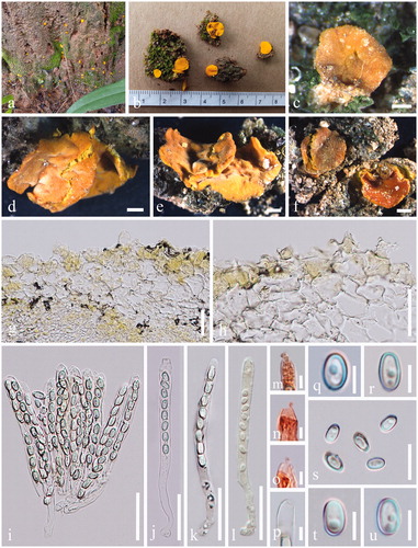
Table 4. Morphological differences for two collections of Acervus globulosus.
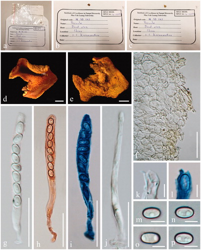
Figure 4. Acervus rufus (HKAS 107302, holotype). (a–c) Typic mature specimens. (d, e) Receptacle surface of pileus. f Hymenium in Congo red. (g) Apex of asci in Congo red. (h) Paraphyses in Congo red. (i–k) Asci (j Asci in Congo red. k Asci in Melzer’s reagent.). (l–o) Ascospores. Scale bars: d–f, i–k = 50 μm. h = 30 μm l = 20 μm. g, m–o = 5 μm.
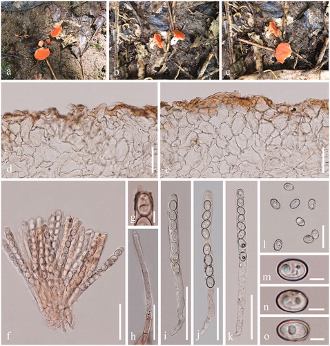
Figure 5. Acervus stipitatus (HKAS 107302). (a) Habitat. (b, c) Typic mature specimens. (d) Receptacle surface of pileus. (e–g) Paraphyses (g Paraphyses in Congo red.). (h–j) Asci (i-i Asci in Congo red.). (k–m) Apexes of asci. (n–q) Ascospores. Scale bars: d, h–j = 30 μm. e–g = 20 μm. k–m, o–q = 5 μm. n = 15 μm.
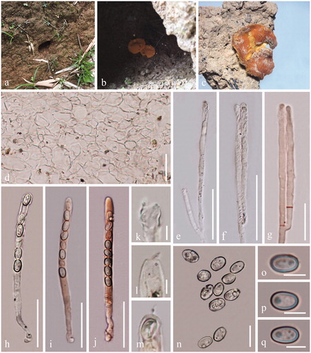
Figure 6. Acervus stipitatus (MFLU 16-0607, holotype). (a–e) Herbarium materials. (f) Receptacle surface of pileus. (g–i) Asci (h Asci in Congo red. i Asci in Cotton blue). (k–l) Apexes of asci. (l Apex of asci in Cotton blue). (m–p) Ascospores. Scale bars: d–i = 30 μm. j–l, n–p = 5 μm. m = 15 μm.