Figures & data
Figure 1. Geographic distribution of Pseudoperonospora cubensis specimens collected on oriental pickling melon (Cucumis melo var. conomon) in Korea. The four plots where P. cubensis specimens were collected are marked on Google Earth map with different colored circles; red for plot 1, blue for plot 2, purple for plot 3, and yellow for plot 4.
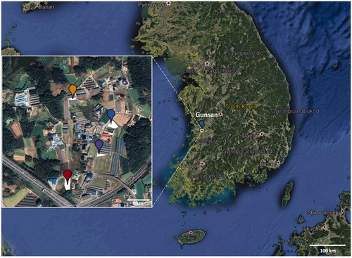
Figure 2. Downy mildew disease caused by Pseudoperonospora cubensis on oriental pickling melon (Cucumis melo var. conomon) in Korea. (A and B) Downy mildew outbreak in a field of oriental pickling melon; (D and E) Vein-limited spots above (D) and below (E) an infected leaf; (G and H) Close-up view of vein-limited downy mildew growth developing on the lower surface; (J and K) Dense sporangiophores with grayish, numerous sporangia; (C and F) Sporangiophore; (I and L) Sporangium. Scale bars: 100 μm for sporangiophore; 10 μm for sporangium.
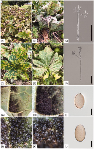
Table 1. Information of Pseudoperonospora collections parasitic to oriental pickling melon (Cucumis melo var. conomon) used in this study.
Figure 3. Minimum evolution tree of Pseudoperonospora species using cox2 mtDNA sequences. Bootstrapping values (minimum evolution BP/maximum likelihood BP) higher than 60% are shown above the branches (10,000 replicates). The scale bar equals the number of nucleotide substitutions per site. Pseudoperonospora specimens collected in four plots were marked as different colors; red for plot 1, blue for plot 2, purple for plot 3, and yellow for plot 4.
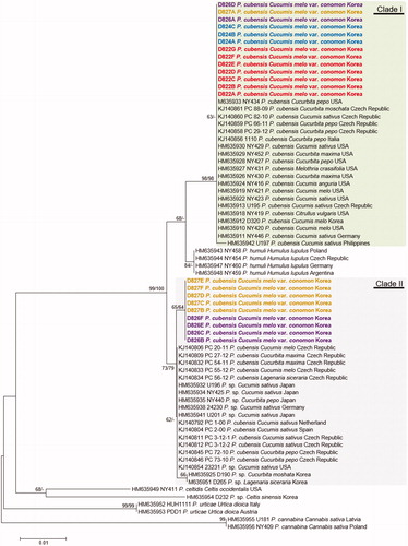
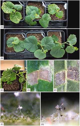
Figure 4. Pathogenicity assay of Pseudoperonospora cubensis isolate D826A on oriental pickling melon (Cucumis melo var. conomon). (A) Oriental pickling melon plants a week after inoculation; (B) Uninoculated plants a week after inoculation; (C–E) Vein-limited spots above (C and D) and below (E) an infected leaf; (F and G) Sporangiophores emerging from stomata, with immature sporangia without melanin pigment (F) and mature sporangia with melanin pigment (G).