Figures & data
Table 1. List of Japanese Botryosphaeria isolates used in this study.
Table 2. PCR primer sets and annealing temperatures.
Table 3. List of Botryosphaeria species used for phylogenetic analysis.
Figure 1. Phylogenetic tree of Botryosphaeria spp. constructed by ML using the combined ITS, RPB2, TEF1-α, and TUB2 gene sequence datasets. ML bootstrap values and Bayesian PPs are given near the branches (BS/PP). Ex-type strains are in boldface.
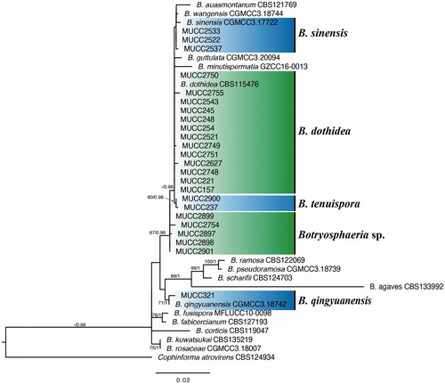
Table 4. Morphological characteristics of the genus Botryosphaeria.
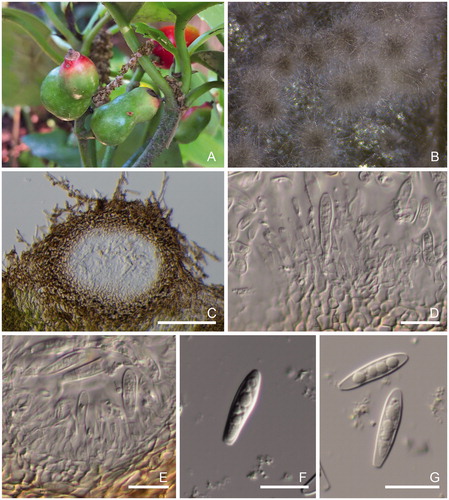
Figure 2. Morphological features of Botryosphaeria tenuispora [A–F: MUMH 10420 (MUCC 237) and G–I: MUCC 2900]. (A) Specimen MUMH 10420. (B) Symptoms with pycnidia forming on the leaf of Leucothoe fontanesiana. (C) Vertical section of pycnidium in the leaf tissue. (D) Conidia and conidiophores. (E, F) Conidia. (G) Conidiomata formation on BMA after 7 days. (H) Conidiomata. (I) Conidium and conidiophores. (J) Conidium. Scale bars, 200 μm (C and H) and 25 μm (D–F and I–J).
![Figure 2. Morphological features of Botryosphaeria tenuispora [A–F: MUMH 10420 (MUCC 237) and G–I: MUCC 2900]. (A) Specimen MUMH 10420. (B) Symptoms with pycnidia forming on the leaf of Leucothoe fontanesiana. (C) Vertical section of pycnidium in the leaf tissue. (D) Conidia and conidiophores. (E, F) Conidia. (G) Conidiomata formation on BMA after 7 days. (H) Conidiomata. (I) Conidium and conidiophores. (J) Conidium. Scale bars, 200 μm (C and H) and 25 μm (D–F and I–J).](/cms/asset/d9effbc6-f09a-4665-b7bb-96de691fffaa/tmyb_a_1895486_f0002_c.jpg)
Figure 3. Morphological features of Botryoshaeria sp. (A and B: MUCC 2899). (A) fruit galls by Asphondylia aucabae on Aucuba japonica. (B) Conidiomata formation on the BMA after 7 d. (C) Vertical section of pycnidium in the leaf tissue. (D–E) Conidia and conidiophores. (F–G) Conidia. Scale bars, 200 μm (C) and 25 μm (D–G).