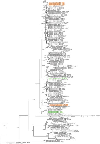
1,197
Views
0
CrossRef citations to date
0
Altmetric
Research Articles

Diversity of the Bambusicolous Fungus Apiospora in Korea: Discovery of New Apiospora Species

, , , , , & ORCID Icon show all
Pages 302-316 | Received 24 Aug 2022, Accepted 05 Oct 2022, Published online: 28 Oct 2022

Figures & data

Figure 1. ML tree based on ITS, LSU, TEF, and TUB concatenated datasets. The node numbers indicate the Bayesian posterior probabilities (PP) > 0.70 and ML bootstrap support (BS) > 70% as PP/BS. The novel Apiospora cultures examined in this study are shown in bold-face orange color. The unrecorded species are denoted by a green color. Type materials indicated by “T”.

Figure 1. ML tree based on ITS, LSU, TEF, and TUB concatenated datasets. The node numbers indicate the Bayesian posterior probabilities (PP) > 0.70 and ML bootstrap support (BS) > 70% as PP/BS. The novel Apiospora cultures examined in this study are shown in bold-face orange color. The unrecorded species are denoted by a green color. Type materials indicated by “T”.

Table 1. Strain informations included in the phylogenetic analyses.

Figure 2. Apiospora lageniformis (KUC21686). (A) PDA; (B) MEA; (C) OA; (D, E) conidiogenous cell with conidia; (F) conidia; (G) lageniform conidiogenous cell; (H, I.) clustered conidia under UHR-SEM.

Figure 2. Apiospora lageniformis (KUC21686). (A) PDA; (B) MEA; (C) OA; (D, E) conidiogenous cell with conidia; (F) conidia; (G) lageniform conidiogenous cell; (H, I.) clustered conidia under UHR-SEM.

Figure 3. Apiospora pseudohyphopodii (KUC21680). (A) PDA; (B) MEA; (C) OA; (D, E) conidiogenous cell with conidia; (F) conidia generated on WA medium under light microscope; (G) lobed hyphopodia; (H) conidiogenous cell with ellipsoidal conidia under UHR-SEM; (I) lobed hyphopodia under UHR-SEM.

Figure 3. Apiospora pseudohyphopodii (KUC21680). (A) PDA; (B) MEA; (C) OA; (D, E) conidiogenous cell with conidia; (F) conidia generated on WA medium under light microscope; (G) lobed hyphopodia; (H) conidiogenous cell with ellipsoidal conidia under UHR-SEM; (I) lobed hyphopodia under UHR-SEM.

Figure 4. Apiospora hysterina (KUC21437). (A) PDA; (B) MEA; (C) OA; (D) conidia; (E, G) conidiogenous cell with conidia; (F, H, I) conidia under UHR-SEM.

Figure 4. Apiospora hysterina (KUC21437). (A) PDA; (B) MEA; (C) OA; (D) conidia; (E, G) conidiogenous cell with conidia; (F, H, I) conidia under UHR-SEM.

Figure 5. Apiospora paraphaeosperma (KUC21488). (A) PDA; (B) MEA; (C) OA; (D, E) conidiogenous cell with conidia; (F) conidia generated on WA medium under light microscope; (G–I) conidiogenous cell with conidia under UHR-SEM.

Figure 5. Apiospora paraphaeosperma (KUC21488). (A) PDA; (B) MEA; (C) OA; (D, E) conidiogenous cell with conidia; (F) conidia generated on WA medium under light microscope; (G–I) conidiogenous cell with conidia under UHR-SEM.

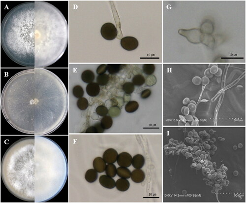
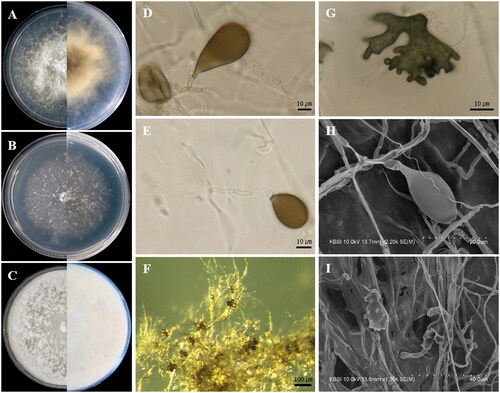
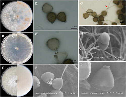
Table 2. List of bambusicolous Apiospora in worldwide.

Supplemental material

Supplemental Material

Download MS Word (220.2 KB)