Figures & data
Figure 1. Morphological characteristics of Miracula polaris. (A) Early infection stages until zoospore maturation; (B, C) late stages of infection: (B) formation of primary cysts; (C) zoospore release; (D) empty cysts (asterisk) in the host cell after the release of secondary zoospores. The scale bar equals 50 µm in all pictures.
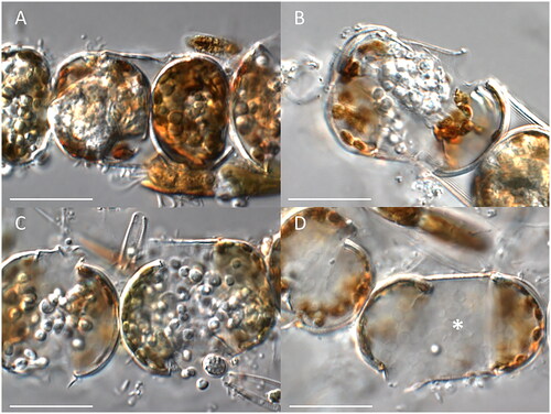
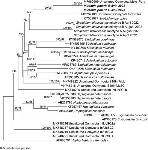
Figure 2. Phylogenetic reconstruction in minimum evolution based on partial nrSSU sequences. Numbers near nodes represent bootstrap support values in minimum evolution and maximum likelihood. A minus sign denotes lack of support for the presented or an alternate topology.
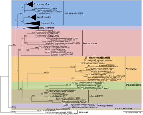
Figure 3. Phylogenetic reconstruction in minimum evolution based on partial cox2 sequences. Numbers near nodes represent bootstrap support values in minimum evolution and maximum likelihood. A minus sign denotes lack of support for the presented or an alternate topology.