Figures & data
Table 1. The antifungal activity of 3-hydrazinoquinoxaline-2-thiol was evaluated using a broth microdilution assay and compared to that of Amphotericin B.
Figure 1. An extensive visual examination was conducted to analyze the standard lesions observed on the tongues of mice afflicted with oral candidiasis. These lesions are typically characterized by the presence of white patches, indicative of fungal infection. The examination involved careful scrutiny of each mouse’s tongue under standardized conditions to distinguish between two distinct states: (A) tongues exhibiting a normal appearance without any discernible abnormalities, and (B) tongues displaying the characteristic white patches associated with oral candidiasis. This meticulous observation process allowed for the detailed characterization and differentiation of the two tongue conditions, facilitating a comprehensive assessment of the extent of fungal infection in the experimental mice.
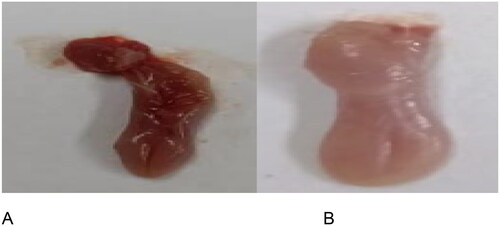
Figure 2. The in vivo effectiveness of 3-hydrazinoquinoxaline-2-thiol using mice model against Candida albicans ATCC 10231. When 3-hydrazinoquinoxaline-2-thiol was administered to mice at various concentrations (0.002, 0.02, and 0.2 mg/ml), we noted a notable decrease in the levels of various inflammatory markers in comparison to the infected mice group that did not receive any treatment. This reduction was statistically significant, with a p-value below 0.05. G1 represents group received 0.002 mg/ml, G2 represents group received 0.02 mg/ml, G3 group received 0.2 mg/ml. (A) TNF-A, (B) IL6, (C) IL-B1, (D) Cox2, (E) INOs, (F) IFN-Y. The levels of TNF-A, IL6, IL-B1, Cox2, iNOS, and IFN-Y were measured in picograms per milliliter (pg/ml) for each respective inflammatory marker.
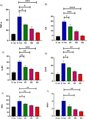
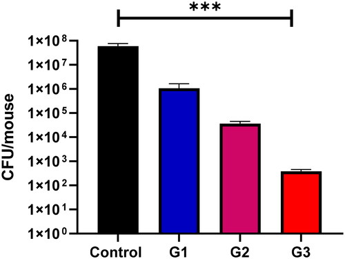
Figure 3. In the context of mouse oral infections, at day 7, the group that received no treatment showed the highest colony-forming unit (CFU) counts. Conversely, the group treated with 0.5 mL of 3-hydrazinoquinoxaline-2-thiol at a concentration of 0.2 mg/ml exhibited the lowest CFU counts. A notable and statistically significant difference was observed between the control group and the treated groups of mice, with a p-value less than 0.05. Moreover, the CFU counts in the treated group decreased as the concentration of 3-hydrazinoquinoxaline-2-thiol increased.