Figures & data
Table 1. Experimental design of the study.
Table 2. Effect of RtH on the survival rate of experimental mice supplemented with different doses of RtH for five consecutive days before exposure to gamma irradiation with a dose of 7.5 Gy. The experimental groups are as described in .
Figure 1. Spleen colony assay on day 11 post-irradiation. The experimental groups are designated as described in .
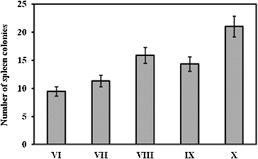
Figure 2. Survival curves of mice supplemented with different doses of RtH: 50 mg/kg (A), 100 mg/kg (B), 150 mg/kg (C) and 200 mg/kg (D). The experimental groups are designated as described in . Mice were irradiated with 7.5 Gy gamma radiation. Survival was monitored up to 30 days post-irradiation. Statistically significant difference between Group VI (irradiated only), and irradiated Groups supplemented with RtH ( p < 0.05).
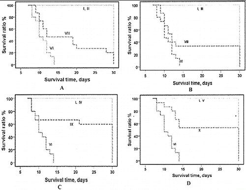
Figure 3. Histological haematoxylin/eosin (HE) analysis of experimental gastric ulcers induced by gamma irradiation. Stomach of healthy mice (A), HE × 10; nerve endings in the underlying tissue of the stomach in healthy mice (B), HE × 40; ulceration on the 4th hour after irradiation (C) – noduli lymphatici (thin arrow) and dilated vessel (thick arrow), HE × 40; and vacuolization (D) of the stomach epithelium (thin arrow) and area of normal epithelium (thick arrow), HE × 40; stomach integrity on the 12th hour (E) and 24th hour (F) after radiation exposure; HE × 40.
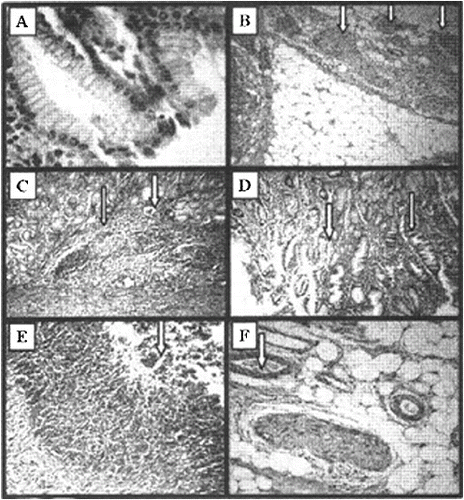
Figure 4. Stomach ulcer area (mm2) of mice 4–24 h after irradiation exposure. The experimental groups are designated as described in . Statistical significance: *** p < 0.001 vs. Group I; +++ p < 0.001 vs. Group VI; ++ p < 0.01 vs. Group VI; n.s. – non-significant.
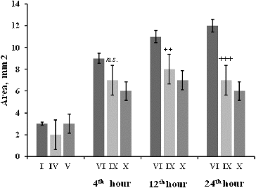
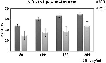
Figure 5. Antioxidant activity (AOA) of RtH and BHT in liposomal syspension. Data are expressed as mean ± SE.