Figures & data
Figure 1. Water suspensions of the studied detonation nanodiamonds with concentration 0.1 mg/ml.
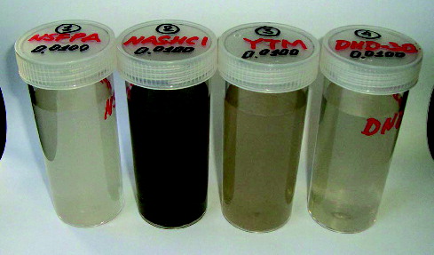
Figure 2. Size distribution of DND particles.
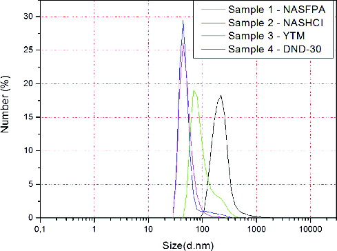
Table 1. ICP-MS analysis of DND samples.
Figure 3. Proliferation activity of MG-63 cells (A) and rMSCs (B), incubated for three days with different DND particles.
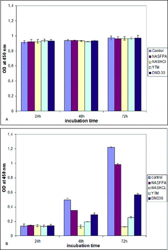
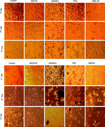
Figure 4. Overall morphology of MG-63 (A) and rMSCs (B) cells, incubated for three days with different DND particles, bar = 100 μm.